Computer-aided detection (CAD) of digital chest tomosynthesis studies helped improve the diagnostic performance of inexperienced readers in a study by Japanese researchers published December 13 in Radiology. But CAD's effect was not statistically significant with physicians who had more experience.
In a study of 100 patients, the use of CAD to analyze suspicious lung nodules found on digital tomosynthesis studies helped improve the overall performance of a group of 15 readers by three points. But CAD's effect varied based on experience level, with interns and residents getting a statistically significant eight-point boost while more experienced readers saw less of an impact (Radiology, December 13, 2017).
"CAD significantly improved diagnostic performance in the detection of pulmonary nodules on chest tomosynthesis images for interns and residents, but provided minimal benefit for chest radiologists and abdominal radiologists," wrote a team led by Dr. Yoshitake Yamada, PhD, from Keio University.
Why not tomosynthesis?
While tomosynthesis is best known for its growing use in breast imaging, proponents believe it could find a role as a sort of intermediary technology between digital radiography (DR) and CT. Tomosynthesis avoids the problem of overlapping anatomical structures that can bedevil radiography, but it is cheaper and had a lower radiation dose than CT.
Meanwhile, CAD algorithms have been developed for a wide variety of clinical applications, ranging from digital mammograms to chest exams with both DR and CT. But there has been little research in the use of CAD for assessing pulmonary nodules found with digital tomosynthesis, according to Yamada.
The researchers therefore wanted to see how well CAD would help a group of readers of varying experience levels interpret suspicious lung nodules found on chest tomosynthesis studies. They performed digital tomo and CT exams on a group of 100 patients who were scheduled for exams to evaluate or screen for lung malignancies from December 2012 to June 2013. Fifty of the patients ended up having pulmonary nodules, while 50 did not.
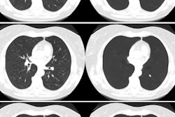
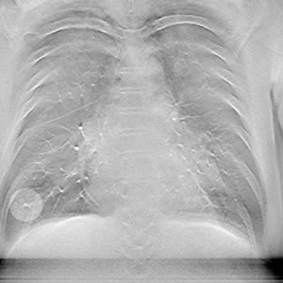
 CAD highlights suspicious lung nodule finding on digital tomosynthesis slice. Image courtesy of Dr. Yoshitake Yamada.
The DR system used was a radiography/fluoroscopy tilt table (Sonialvision Safire, Shimadzu Medical Systems) that was tilted to 80° to reduce motion artifacts as patients leaned back against the table. The system acquired 41 coronal images as the x-ray tube moved around the patient in an arc of 20° over five seconds. The average radiation dose to patients was 0.19 mSv. The CT studies used a standard chest imaging protocol, with an effective dose estimate of 2 mSv to 3 mSv.
The tomosynthesis studies were analyzed with commercially available software (Cadviser TS, Shimadzu) that is optimized to detect lung nodules that are 5 mm to 20 mm in diameter and width (nodules that are smaller and larger than this range are also marked, the authors noted).
Finally, both the tomosynthesis and CT studies were analyzed by a group of 15 readers of varying experience levels. Five were interns or residents, five were board-certified specialty-trained chest radiologists, and five were board-certified subspecialty-trained abdominal radiologists. The CT scans were considered to be the reference standard for nodule detection.
Readers were instructed to search the tomosynthesis images first without CAD, and then again with CAD marks displayed. They were allowed to dismiss the markings that the CAD algorithm thought might be nodules.
How they measured up
Yamada and colleagues analyzed the diagnostic performance of readers with and without CAD using free-response receiver operating characteristic (ROC) analysis, generating figures-of-merit (FOMs) for the performance of the readers with and without CAD. They then classified the scores by experience level.
Analyzing the data by the entire group, the researchers found that CAD contributed to a slight improvement in overall performance that was statistically significant. But the gains were concentrated among the inexperienced readers versus more experienced radiologists, as indicated in the table below.
| Effect of CAD tomosynthesis on lung nodule detection | |||
| ROC before CAD | ROC after CAD | p-value | |
| Overall | 0.71 | 0.74 | 0.02 |
| Chest radiologists | 0.78 | 0.80 | 0.38 |
| Abdominal radiologists | 0.73 | 0.74 | 0.65 |
| Interns and residents | 0.62 | 0.70 | 0.001 |
The researchers said their results were in line with a previous study that found an increase in nodule detection for inexperienced readers who underwent training feedback on tomosynthesis; there was no similar benefit for experienced readers, however.
"We found that CAD was especially beneficial for interns and residents, but minimally beneficial for trained radiologists," they wrote. "Our study is similar to that previous study in that inexperienced readers would require training or additional assistance, such as that provided by CAD, whereas experienced readers may not need this assistance."
In secondary findings, the researchers noted that the CAD algorithm had a false-positive rate of 3.22 for all cases, which they said "may still be unsatisfactory." On the other hand, the actual average false-positive rate for all cases with human assessment was only 0.26, which was lower than the CAD rate, indicating that the readers were taking the CAD marks with a grain of salt.
"Presumably, this [false-positive rate] was because the observers disregarded CAD-detected nodules appropriately," they concluded.