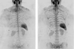
Low-dose whole-body CT detected almost four times as many lesions as the gold standard, radiographic skeletal survey (RSS), in multiple myeloma patients, researchers reported on Wednesday at the American Roentgen Ray Society (ARRS) meeting in Vancouver.
The use of CT for multiple myeloma diagnosis is gaining favor in Europe, but it has not been extensively studied in U.S. patients, where radiography is the norm, according to Dr. Kelechi Princewill and colleagues from the University of Maryland. Skeletal surveys do a good job of detecting lesions in the appendicular skeleton and skull, but they can be less accurate for detecting lesions in the axial section, according to the group.
The authors retrospectively reviewed 300 multiple myeloma cases, spanning 13 years, in which patients were diagnosed using both RSS and PET/CT; only the low-dose CT component of PET/CT was used for comparison.
The low-dose CT study delivered an average dose of 4.1 mSv, compared with 1.8 mSv for skeletal surveys. The CT average was about nine times lower than doses used in the acquisition of standard body CT studies, the team wrote.
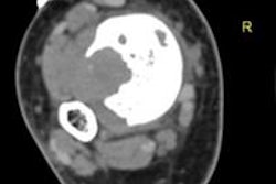
Two radiologists working independently assessed lytic lesions in the skull, spine, sternum, flat bones, and proximal long bones in scans acquired with both modalities in 51 patients (mean age, 56 years).
Whole-body CT found 968 lesions versus 248 for RSS (p < 0.001) in 42 patients, 39 of whom had more lesions at CT than at skeletal survey. Eight cases with lesions at CT would have been missed.
In 31 patients (61%), the number of lesions found on CT would have resulted in upstaging of the disease. Thirteen patients would have been upstaged from stage I to II, nine from stage I to III, and nine from stage II to III.