Another milestone has been reached in establishing equivalency between digital radiography (DR) and conventional film-screen chest radiography for characterizing pneumoconiosis, a set of pulmonary diseases associated with job-related black lung disease and asbestosis.
In the latest study in a series of trials, researchers found no statistically significant difference between the characterization of pleural abnormalities on DR versus film-screen radiography. The research supports a recent initiative by the U.S. Department of Health and Human Services to allow lung studies of pneumoconiosis to be performed with digital x-ray systems in addition to traditional film-based radiography.
Theodore Larson, an epidemiologist with the U.S. Agency for Toxic Substances and Disease Registry, was the lead author of the study, which was published in Academic Radiology (February 2012, Vol. 19:2, pp. 131-140).
A major step
The conversion from film to digital radiography is a major step for physicians and epidemiologists, who oversee the international system that has been used to detect and monitor pneumoconiosis since the 1930s. The system has helped establish a scientific basis for coal mine regulation, litigation, and individual worker compensation claims.
Worldwide, radiographs for characterizing pneumoconiosis are coded using International Labour Organization (ILO) classification. This requires comparing a patient's chest film with a set of 22 standard film-screen radiographs that describe the various shape, size, and severity of abnormalities found in pneumoconiosis patients. Physicians undergo special training for certification to read the scans.
The ILO system still requires the use of analog films, despite the ongoing shift to digital x-ray. This transition has created the need to validate digital x-ray for classifying pneumoconiosis, according to the authors. A few such studies have been conducted, but these have focused on parenchymal abnormalities, which correlate with coal and silica dust exposures that contribute to pneumoconiosis and silicosis.
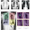
In the current study, Larson and colleagues focused on pleural lung abnormalities, which are often associated with asbestos exposure and asbestosis. The researchers performed DR and film-screen x-ray chest studies on 200 patients exposed to asbestos.
The studies were read independently twice by seven readers using conventional ILO standards for film and digitized standards for digital imaging. Areas under the receiver operator characteristics (ROC) curve were calculated using chest CT scans as the gold standard. The CT scans were interpreted by three readers.
Overall image quality was high for film-screen and DR images for ILO classification. Good image quality was attained for 61.6% of the film-screen images and 66% of the DR images. Another 36.9% of the film-screen and 32.6% of the DR images were rated as acceptable, with no technical defect. No image from either approach was deemed nondiagnostic.
Though differences were not statistically significant, film-screen x-ray was slightly better than DR for measuring the prevalence of pleural abnormalities (56.2% versus 54.3%).
"We found that the difference between film and digital radiography for classifying pleural plaque was very small and not statistically significant," Larson said in an interview. "In practical terms, we did not find a reason for not using digital for the purposes of classifying pleural abnormalities."
Larson's study fills a gap in the growing medical literature on the equivalence of DR and film-screen because of its emphasis on pleural abnormalities, epidemiologist A. Scott Laney, PhD, told AuntMinnie.com.
Laney, who works for the U.S. National Institute for Occupational Safety and Health (NIOSH), prepared one of several published studies that have established equivalency between analog x-ray and computed radiography and DR for classifying parenchymal lung abnormalities. Though minor questions remain, epidemiologists are now satisfied that DR and film-screen imaging are equivalent for characterizing pneumoconiotic abnormalities, Laney said.
Larson, however, supports a final formal evaluation of all equivalency studies to determine if the weight of evidence permits the formal adoption of DR for use with the ILO classification system.