Digital tomosynthesis may help assess pulmonary tuberculosis (TB) more thoroughly than general radiography while serving as a low-dose alternative to multislice CT, the traditional modality of choice when imaging is required to monitor the highly contagious disease.
A new low-dose tomo protocol, developed at Samsung Medical Center in Seoul, South Korea, is proving more sensitive to the presence of TB-related lung abnormalities than conventional digital radiography (DR) without exposing patients to much more ionizing radiation.
One-third of the world's population is thought to be infected with the Mycobacterium tuberculosis bacterium that causes TB. Infection does not necessarily lead to illness: The body's immune system can encapsulate the bacilli, allowing them to lie dormant for years.
A chest radiograph to exclude pulmonary TB is used to select patients for latent tuberculosis infection treatment. Cavities, nodules, and fibrotic scars detected with radiography may contain replicating tubercle bacilli that could lead to active TB, according to the World Health Organization. Calcified nodular lesions and apical pleural thickening signify a lower risk for progression. Some pulmonary infiltrates may only be confidently characterized with CT.
In an observer study presented at the 2009 RSNA meeting in Chicago, Drs. Myung Jin Chung and Kyung Soo Lee discussed how a low-dose digital tomosynthesis protocol developed in their lab outperforms digital radiography and approximates the sensitivity of 64-slice CT for detecting diverse parenchymal lung lesions, including cavities associated with pulmonary TB.
The protocol was designed using a commercially available DR system (Definium 8000, GE Healthcare, Chalfont St. Giles, U.K.) equipped with the company's VolumeRAD tomosynthesis module. The module acquires 3D volumes by acquiring data while the x-ray tube moves from a negative 17.5° to a positive 17.5° arc around the standard posterioanterior position, with 60 low-dose projection images acquired at a tube voltage of 100 kVp during 10 seconds. The data were then reconstructed for presentation in 54 coronal images.
Lee's group established that the low-dose protocol delivered an effective dose of 0.05 mSv, about half the radiation usually involved with tomosynthesis and far less than the 3.4 mSv that is typical during a 64-slice chest CT procedure.
Digital chest radiography was also performed for the trial on the Definium 8000 at 120 kVp under automatic exposure control at a film speed equivalent of 400. This delivered an effective dose of 0.02 mSv. In practice, a two-shot exam enabling bone subtraction and involving anteroposterior and lateral views would expose the patient to an effective dose of 0.07 mSv, according to Chung and Lee.
Sixty-five patients (32 men and 33 women) with pulmonary TB were examined with digital tomo, DR, and CT. Using CT as a gold standard, Lee found that tomosynthesis was significantly more sensitive than radiography for all types of TB-related lung lesions.
 |
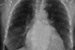
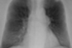
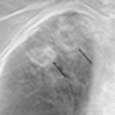
| Two thick-walled cavities from an 80-year-old man with active pulmonary TB were seen in digital tomo image (left). They were confirmed on CT (center) but escaped detection in the radiograph (right). Images provided by Dr. M.J. Chung. |
For the presence of cavities, for example, tomosynthesis had a per-lesion sensitivity of 85%, characterizing 85 of the 100 cavities observed with CT. In comparison, general radiography identified 28 cavities, for a per-lesion sensitivity of 28%. Both modalities were responsible for three false-positive lesions in two patients.
Per-patient sensitivity of digital tomo and digital radiography for TB-related lung lesions
|
The researchers also found a high level of agreement between the two radiologists interpreting the digital tomo scans compared to radiography. The mean kappa value was 0.90 for tomo compared to 0.68 for radiography.
In a written response to questions, Chung and Lee were optimistic that their group's low-dose technique will increase the acceptance of tomo for detecting diverse lung lesions.
"We anticipate the alternative use of low-dose digital tomosynthesis for evaluation of patients with pulmonary tuberculosis, especially for the follow-up or monitoring after treatment of tuberculosis," they wrote.
Other research by the group suggests that low-dose digital tomo will also outperform radiography for many nonpulmonary clinical applications.
Study results were limited by the readers' lack of experience interpreting digital tomo images and the decision to not gauge disease activity using clinical data from sputum smears and physical symptoms.
By James Brice
AuntMinnie.com contributing writer
March 19, 2010
Related Reading
Virtual dual-energy DR improves lung lesion detection, February 10, 2010
Rads prefer dual-energy x-ray over standard DR in lung, January 29, 2009
Adding CsI-based dual-energy imaging doesn't improve lung nodule detection, December 26, 2008
Dual-energy digital x-ray still looking for acceptance, February 22, 2007
Copyright © 2010 AuntMinnie.com