Researchers from the University of Washington have found that digital radiography (DR) tomosynthesis can help radiologists assess the healing process in patients with wrist fractures -- especially those with metal hardware that might confound CT scans.
In a study presented at last week's American Roentgen Ray Society (ARRS) meeting in San Diego, Dr. Alice Ha from the university's department of radiology discussed her group's effort to find a better tool than radiography for following up patients who have fractured their wrist.
Tomosynthesis not only performed nearly as well as CT, the gold standard, it did so at a radiation dose only slightly higher than that of conventional radiography, she said. Ha and colleagues have developed a clinical algorithm to integrate tomosynthesis into everyday practice.
A tricky target
Radiography is typically used to follow up patients with wrist fractures, allowing physicians to monitor the healing process and give patients the go-ahead to return to normal activities. Imaging can also be used to direct patients to surgery in more severe cases of fracture.
 Dr. Alice Ha from the University of Washington.
The wrist presents a tricky target for plain x-ray, however. The multiple bones in the wrist can overlap, making it difficult to view pathology on 2D radiographs. And metal implants that are increasingly being used to help the healing process can also create problems.
CT isn't typically used for monitoring wrist fractures due to its higher radiation dose and susceptibility to artifacts, although physicians will use it for problem-solving, Ha said. Therefore, a better technology is needed.
On the advice of Dr. Felix Chew, section head of musculoskeletal radiology at the university, Ha investigated the tomosynthesis module that had been installed on one of the department's DR systems (VolumeRad, Definium 8000, GE Healthcare).
Ha's team decided to compare tomosynthesis to radiography in patients being treated for wrist fractures at the university. Patients received both tomo and conventional radiography within one year of hardware placement, with the tomosynthesis system acquiring 60 slices. Noncontrast CT scans were also acquired to create a gold standard for comparison.
Three readers evaluated the tomosynthesis and radiography images, assessing them in two main areas: fracture healing and complications from the hardware placement. Reader performance was evaluated using receiver operator characteristics (ROC) analysis, with the area under the curve (AUC) used to assess each modality against CT.
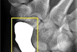
The researchers evaluated a total of 49 patients (31 men, mean age of 47 years) with a total of 51 bones with implanted hardware. The most common indications were distal radius fracture (43%), scaphoid fracture (18%), and metacarpal fracture (18%), and fracture lines were analyzed for four cortices -- medial, lateral, anterior, and posterior.
DR tomosynthesis performed significantly better than conventional radiography, the group found. Tomo had an AUC of 0.84 for detecting cortical fracture lines, compared with 0.76 for radiography (p = 0.01).
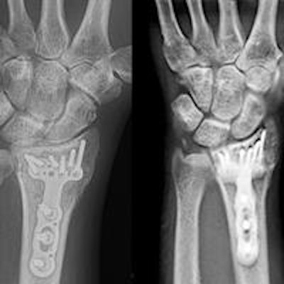
 At left, frontal radiograph of a 41-year-old woman with healing comminuted distal radial fracture, post internal fixation. At right, coronal reconstruction image from tomosynthesis. Images courtesy of Dr. Alice Ha.
Regarding complications from hardware placement, cortex obscuration occurred in 2% of CT images, 8% of tomosynthesis images, and 15% of radiography images (p < 0.01 between each modality).
Finally, the researchers tried to correlate the patients' fracture scores with their clinical outcome as measured by pain and Disabilities of the Arm, Shoulder, and Hand (DASH) scores, but they found no significant correlation. There was a statistically significant correlation between pain levels and fracture scores for tomosynthesis (r = 0.28, p = 0.03) and CT (r = 0.29, p = 0.04).
With respect to radiation, Ha and colleagues investigated previously published research on tomosynthesis and also performed studies on cadavers. They found that the radiation dose from tomo is comparable to radiography, she said.
In future research, Ha's team hopes to assess tomosynthesis for visualizing weight-bearing feet and ankles. They have also developed protocols for using tomosynthesis in clinical practice, most likely in conjunction with two-view radiography.
"There was some learning curve that had to be overcome by the readers -- they had never looked at tomosynthesis before -- but once they got their eyes adjusted, in the end everyone liked it," she said.