Real-time elasticity imaging (EI) improves the characterization of breast lesions, according to the results of a multicenter study presented at the 2007 RSNA meeting in Chicago. The researchers noted that malignant lesions appeared larger in size and volume than benign lesions on the elastograms.
"Elasticity generates images based on the stiffness or hardness of a lesion and correlates to a physical examination, but with imaging," explained Dr. Richard Barr, Ph.D., from Radiology Consultants in Campbell, OH, and St. Elizabeth Health Center in Youngstown, OH. "This technique is now commercially available around the world."
Barr, who is the principal investigator for the trial to determine the sensitivity and specificity of EI in breast lesions, presented an update on his group's work at this year's meeting. At the 2006 RSNA meeting, he discussed the results of a single-institution, pilot study done in 80 women (123 breast lesions), all of whom underwent biopsy. The group found EI had 100% sensitivity and 98% specificity for identifying cancers.
"The aim of this (current) study was to see if we could reproduce the results from the one-center site with other sites among people who were just trained in the technique," Barr said.
 |
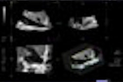
| Biopsy-proven IDC. Above, a conventional ultrasound image. Below, EI acquired at the same time with the same signals. The lesion in the elasticity image is much larger than the image in the conventional ultrasound, which is more likely to correspond to a malignant lesion. Images courtesy of RSNA. |
 |
The 635 women in this arm were scheduled for biopsy for suspected breast lesions and underwent standard imaging plus EI, done on a modified Elegra or Antares ultrasound system and software (Siemens Medical Solutions, Malvern, PA). The elastogram added no more than two minutes to the exam time, Barr said.
The lesions evaluated included infiltrating ductal carcinoma (IDC), ductal carcinoma in situ (DCIS), fibroadenomas, and cystic lesions. The average lesion size was 10 mm. Side-by-side, simultaneous sonographic images of the breast were produced in B-mode and with EI. The radiologist at each site measured the lesions and classified them as benign or malignant. Biopsy and its pathological results served as the reference standard.
According to the results, 137 of 139 malignant lesions had an EI-to-B-mode ratio of 1.0 or greater. Among the 245 benign lesions, 199 had an EI-to-B-mode ratio of less than 1.0. Once again, EI turned in a high sensitivity: 100% at five of six sites and 97% at the sixth one. There were 413 biopsy-proven lesions among the women studied, Barr said, and 222 were biopsy-proven malignancies.
In addition to differences in shading compared to normal tissue, EI depicted lesion stiffness or hardness, as well as changes in size. "Malignant lesions appear to be larger on the elastogram, while benign lesions appear to be smaller," Barr stated.
Although the overall specificity was decent at 81%, specificity across the individual sites ranged from 96% to 48%. Barr and colleagues attributed this variability to operator dependence.
"There is a learning curve with this technique," Barr said, adding that the amount of pressure placed on the transducer can affect EI outcome, with a lighter touch producing better-quality images.
By Edward Susman
AuntMinnie.com contributing writer
December 14, 2007
Related Reading
New techniques in breast ultrasound may find more cancers, March 1, 2007
US elasticity testing could cut breast biopsies by half, November 28, 2006
Streaming US, elastography US give clearer picture of breast lesions, June 16, 2006
Copyright © 2007 AuntMinnie.com