Computer-aided detection (CAD) took a hit last month when a study in the New England Journal of Medicine found that the technology's false-positive rate had a negative impact on the accuracy of mammography screening. But two recent studies indicate that CAD still produces beneficial results when paired with a couple of new breast screening technologies, CR mammography and full-field digital mammography.
One of the studies backing a more beneficial view of CAD comes from the George Washington University Medical Center in Washington, DC, which reviewed cases from clinical trials at two sites to evaluate CAD's performance with CR mammography in detecting breast cancer. The evaluation criteria included breast density, mammographic appearance, histopathology, and lesion size.
Both sites utilized the FCRm CR mammography system from Fujifilm Medical Systems USA of Stamford, CT. The CAD technology for the study was SecondLook version 7.2 from iCAD of Nashua, NH, which is being used on an investigational basis with CR mammography. iCAD vice president and medical director Dr. Jeff Hoffmeister presented the results at the 2007 American Roentgen Ray Society (ARRS) meeting earlier this month.
CAD-CR data
Researchers evaluated CAD's performance on 53 breast cancers detected with CR mammography. The cases included mammographically detected lesions, BI-RADS 4 or 5 lesions, malignancies detected through biopsy, and craniocaudal (CC) and mediolateral oblique (MLO) mammograms from the breast with the lesions.
Thirty-six normal cases were used to determine the system false-positive (FP) rate. Those cases were interpreted as BI-RADS 1, with four standard views, both as CC and MLO.
The study's radiologists assessed breast density of malignant and normal mammograms; mammographic appearance of cancers, whether they were microcalcifications and masses, such as spiculated and nonspiculated masses, architectural distortions, and/or focal asymmetries; and the location of cancers on the mammograms.
 |
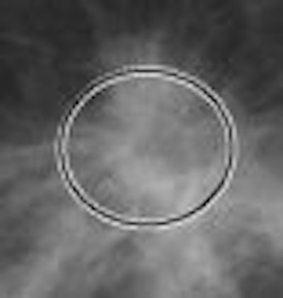
| Microcalcifications (above) within the rectangular frames show the extent the calcifications that are detected by CAD. Masses (below), architectural distortions, and focal asymmetries are marked with the circular frame. Images courtesy of iCAD. |
 |
Researchers determined that 47 of 53 cancer cases were detected by the CAD system, resulting in a sensitivity of 89%. In addition, 30 of 33 cancers (91% sensitivity) were detected in nondense breasts, while 17 of 20 cancers (85% sensitivity) in dense breasts were discovered.
The CAD system's false-positive rate was 2.2 marks per four-image case.
In addition, the CAD technology detected 92% of cancers (11 of 12 cases) manifested as calcifications and 88% (36 of 41 cases) as masses. Twenty-two masses were spiculated, of which CAD correctly marked 20 masses for a sensitivity of 91%.
For cases of invasive ductal carcinoma, the detection rate was 89% (31 of 35 cases); invasive lobular and other carcinomas was 80% (four of five cases); and ductal carcinoma in situ was 92% (12 of 13 cases).
CAD's sensitivity for cancers 1 mm to 10 mm was 83% (15 of 18); 11 mm to 20 mm was 88% (15 of 17); 21 mm to 30 mm was 92% (11 of 12); and greater than 30 mm was 100% (six of six).
In summary, CAD had a sensitivity of 89% with CR mammography that was maintained even in conditions that may lower the sensitivity of mammography -- 85% sensitivity with dense breasts and 83% sensitivity for lesions of 1 cm or smaller.
"We also show that the sensitivity of CAD with CR mammography in spiculated masses (was) 91% and (in) calcifications (was) 92%," Hoffmeister said. "So, we felt that CAD has a high sensitivity for the detection of breast cancer on CR mammography and may result in the earlier detection of breast cancer."
CAD, FFDM, and SFM
Hoffmeister also provided the findings of a study by Columbia University Medical Center in New York City that assessed the effectiveness of CAD with full-field digital mammography (FFDM), as well as how well CAD and FFDM compared to CAD with film-screen mammography (FSM).
"Although the effectiveness of CAD with (film-screen) mammography has been extensively reported in scientific literature, there is less available for CAD with full-field digital mammography," Hoffmeister said. "If the results are similar, it may imply that the outcomes of all the studies with CAD with film may have some applications in CAD with digital mammography."
The retrospective study took 45 cases of breast cancer from five sites, with images collected using the Senographe 2000D FFDM system (GE Healthcare, Chalfont St. Giles, U.K.).
For comparison, 899 breast cancer cases detected with FSM were taken from 18 sites. The cases included lesions detected with screening mammography, malignancies discovered through biopsy, and malignant lesions visible on at least one view. In addition, 147 normal cases were rated as BI-RADS 1, with at least three years of follow-up with BI-RADS 1 or 2 mammograms, and four standard views (both craniocaudal and mediolateral oblique projections).
The cases also were evaluated by SecondLook version 7.2 from iCAD. The technology was developed with CAD algorithms designed to adapt to the characteristics of both digital mammography and digitized film to produce similar outcomes.
Statistically similar
Columbia University researchers found that CAD's sensitivity with FFDM was 89%, as the technology detected 40 of 45 cancers. CAD's sensitivity with FSM was 90%, detecting 809 of 899 cancers. "Therefore, the detection rate of CAD with FFDM and (FSM) was the same," Hoffmeister said.
In addition, when evaluating CAD's performance based on mammographic appearance of cancer on FFDM, eight out of eight cases with calcifications were marked by the technology, while the CAD's sensitivity for masses was 86%, or 32 of 37.
Based on mammographic appearance of cancers on FSM, CAD's sensitivity for calcifications was 95%, or 287 cases out of 292. For masses, 532 of 607 cases were discovered, resulting in a sensitivity of 88%. The CAD system's false-positive rate was 1.6 marks per four-image case with FFDM and 2.0 with FSM.
"This study showed that CAD's sensitivity is statistically the same with digital and film mammography, and the false-positive rate is similar," Hoffmeister concluded. "Thus, we believe the results indicate that CAD with digital mammography will enhance a radiologist's effectiveness for detecting breast cancer in a way that is consistent with the enhancement that has been demonstrated for CAD with film mammography."
By Wayne Forrest
AuntMinnie.com staff writer
May 24, 2007
Related Reading
NEJM study pans CAD, draws attention and criticism, April 5, 2007
CAD boosts single reading of mammograms, September 29, 2006
Multimodality breast CAD workstation improves diagnostic performance, August 17, 2006
Pilot study: Entropy-driven CAD zips through vast breast image database, August 2, 2006
CAD interval change analysis boosts mammogram accuracy, July 7, 2006
Copyright © 2007 AuntMinnie.com