FDG-PET/CT can provide "significant prognostic stratification information" at initial staging for patients with locally advanced breast cancer compared to conventional imaging, according to a study published in the January issue of the Journal of Nuclear Medicine.
French researchers found that FDG-PET/CT more accurately showed lesions in the chest, abdomen, and bones in a single imaging session, and it changed treatment management for more than 50% of the patients in the study.
A team led by Dr. David Groheux, PhD, from the department of nuclear medicine at Hôpital Saint-Louis in Paris, prospectively evaluated 117 patients with locally advanced breast cancer over five years.
Patients received conventional imaging, including bone scans, chest radiography, or dedicated CT and abdominopelvic examination by sonography or contrast-enhanced CT, and were staged. They then received an FDG-PET/CT scan, which was reviewed by nuclear medicine specialists who were blinded to the conventional imaging results.
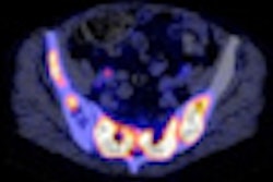
FDG-PET/CT was able to locate all primary tumors, confirming lymph node involvement in stage IIIC patients and revealing unsuspected lymph node involvement in 32 additional patients.
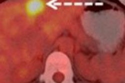
In addition, distant metastases in bone, lymph nodes, liver, lung, and pleura were seen with FDG-PET/CT in 43 patients (37%). Conventional imaging, by comparison, identified 28 patients (24%) with distant metastases.
FDG-PET/CT changed the stage of 61 (52%) of the 117 patients, which affected their recommended treatment.