Using PET/CT to image progesterone-receptor (PR) status may help identify response to endocrine therapy at an early stage and determine the most appropriate treatment for breast cancer patients, according to a preclinical study published in the July issue of the Journal of Nuclear Medicine.
PET has been used to identify the target for endocrine therapy in breast cancer by demonstrating estrogen-receptor (ER) presence with F-18 fluoroestradiol (FES) PET, or by monitoring for hormone-induced changes in tumor metabolism with FDG-PET once therapy has begun, noted the researchers from Mallinckrodt Institute of Radiology at Washington University in St. Louis.
"What is novel about our study is that we chose to image progesterone-receptor levels to see how the estrogen signaling pathway is functioning in response to endocrine therapy," said Dr. Amy Fowler, PhD, lead author of the study, in a statement.
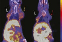
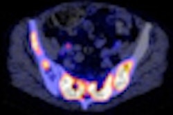
In the study, Fowler and colleagues imaged mice with mammary cell lines (SSM1, SSM2, and SSM3) from tumors that are estrogen-receptor-α (ERα) positive and PR-positive and similar to most human breast cancers. Small-animal PET/CT was performed using FES to image estrogen-receptor status, fluorofuranyl norprogesterone (FFNP) for progesterone-receptor status, and FDG for glucose uptake (JNM, July 2012, Vol. 53:7, pp. 1119-1126).
Initial imaging of the cell lines showed that SSM3 tumors displayed the greatest FES and FFNP uptake. As a result, the SSM3 cell line was used to test response to estradiol, which binds to the estrogen receptor and triggers a response, and fulvestrant, which also which binds to the estrogen receptor but does not prompt a response.
Treatment with estradiol indicated that PR expression is estrogen-inducible and reflects ERα signaling in the SSM3 tumors. Mice treated with fulvestrant showed early decreases in FFNP uptake after therapy and before measurable growth inhibition. SSM2 tumors, which were not inhibited by fulvestrant despite also being ERα-positive/PR-positive, showed no change in FFNP uptake after therapy.
The results, Fowler and colleagues concluded, support the potential use of PR imaging with FFNP-PET of patients with ERα-positive breast cancer at baseline and shortly after initiation of endocrine therapy to distinguish between patients who will and will not respond to therapy.