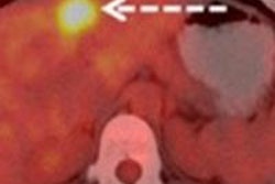
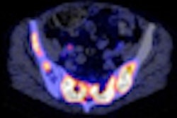
Maximum standardized uptake values on PET/CT scans can help predict prognosis in breast cancer patients with bone metastases, but other PET/CT values could be even more useful, according to research presented last week at the American Society of Clinical Oncology (ASCO) Breast Cancer Symposium.
Dr. Komal Jhaveri and colleagues from Memorial Sloan-Kettering Cancer Center found that in patients with metastatic breast cancer, the standardized uptake value (SUV) of PET/CT correlated with a mortality risk almost three times higher in patients with bone metastases compared to those with lower SUV levels.
"Historically, radiographic prognostication [for metastatic breast cancer] has been very challenging," Jhaveri told attendees at the San Francisco meeting. "But modern PET/CT may be a complementary and clinically useful prognostic tool."
Jhaveri's team identified 285 patients who had received PET/CT scans within 60 days of their diagnosis of metastatic breast cancer, and who had at least one lesion with active F-18 FDG uptake. Tumor activity was measured using SUV uptake.
Lesions detected included bone, liver, lung, and lymph nodes. Due to possible confounding effects of chemotherapy on maximum SUV values (SUVmax), patients who had received chemotherapy within 30 days of the PET/CT exam were excluded, Jhaveri said, leaving 269 patients in the final cohort.
Of the patient group, 87% had invasive ductal carcinoma in situ (DCIS) and 80% had grade 3 cancer, according to Jhaveri. The median time to metastatic breast cancer diagnosis was 2.3 years; 90% of the patient cohort had biopsy-proven metastatic disease and 45% had visceral disease. The most common reason for the PET/CT exam was abnormal radiology results (28%).
For women with bone metastases, the highest tertile of SUVmax carried 2.91-fold higher mortality risk.
"Greater risk of death was seen in patients with higher SUV max in all metastatic sites," she said. "But this was statistically significant only in bone lesions."
SUVmax by site as prognostic variable
|
|||||||||||||||||||||||||||||||||||||||||||||||||||||||||||||||||||||
| Data courtesy of ASCO. | |||||||||||||||||||||||||||||||||||||||||||||||||||||||||||||||||||||
When the researchers conducted a multivariate analysis of the data, they found that the prognostic effect of SUVmax in the bone was maintained when adjusted for grade, tumor phenotype, and presence of visceral metastases -- predicting 2.67-fold mortality risk when SUVmax was high and 1.87-fold risk when intermediate (p = 0.006).
One of the study's limitations was that it didn't evaluate SUVmax as a predictive indicator, but only at one time point, Jhaveri said. She suggested that another way to use PET/CT as a prognostic tool for metastatic breast cancer would be to calculate average SUV across the metastasis rather than the peak SUV in that lesion, a technique called "total lesion glycolysis," or TLG.
"Given the variation in SUV and the differential impact on survival by site, it is possible that SUVmax may not be the most optimal PET variable," she said. "TLG might offer an advantage because it could reflect the density or packing of cells, which relates to histology as well as growth rates and invasiveness of [metastatic breast cancer]."


