Digital breast tomosynthesis (DBT) is an emerging imaging modality in which tomographic images of the breast are generated from multiple 2D x-ray projection images. But DBT can deliver much more -- it also offers the potential for super-resolution (subpixel) imaging.
In a poster presentation at the SPIE Medical Imaging meeting in February, Raymond Acciavatti from the University of Pennsylvania described the latest developments in understanding and optimizing this capability. Acciavatti's work on super-resolution DBT certainly made an impression -- the poster was honored with the conference's Best Student Paper Award.
Proof of principle
DBT works by recording a series of projection images while the x-ray tube is gradually rotated through a small arc with the breast held in compression. As the projection angle changes, the non-normal beam incidence causes the image to be translated in subpixel increments. Reconstructing the overlapping projections creates a 3D image with a resolution that's better than that of the x-ray detector.
To demonstrate DBT's potential to image with super-resolution, Acciavatti and co-author Andrew Maidment, PhD -- chief of physics in the University's radiology department -- used Hologic's Selenia Dimensions digital mammography system to record images of a bar phantom. With 140 µm detector elements, this instrument would be expected to only resolve images up to a frequency of 3.57 line pairs per millimeter (lp/mm).
As expected, central-projection images of bar patterns with 4 and 5 lp/mm exhibited aliasing artifacts, including Moiré patterns, misorientation, and reduced frequency effects. However, tomosynthesis images reconstructed with 11.7-µm voxels could clearly resolve bar patterns with frequencies of up to 6 lp/mm. Reconstruction of the 15 images recorded by the Selenia Dimensions was performed using a commercial back-projection filtering algorithm (Real-Time Tomography).
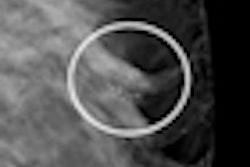
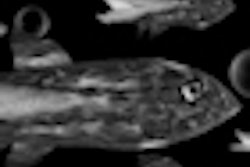
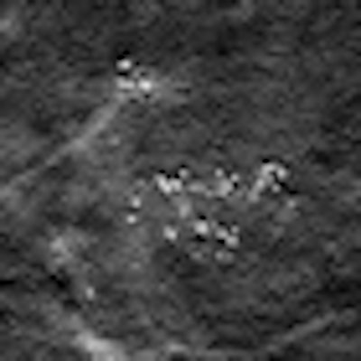
The researchers also compared two breast images taken with the Selenia Dimensions and created in different ways: using voxels that match the detector element size (140 µm); and using a back-projection filtered reconstruction with 35-µm voxels.
 |
| Left: Image created by bilinearly interpolating a conventional reconstruction using 140-µm voxels; the net result has 35-µm voxels. Right: Image created by a back-projection filtered reconstruction with 35-µm voxels; this image displays the microcalcification morphology more clearly. Images courtesy of medicalphysicsweb. |
"You can see microcalcification structural features more clearly in the super-resolution image, and can also see some microcalcifications that you can't see on the other image," Acciavatti explained. "Having a clearer picture of the shape of a microcalcification provides useful diagnostic information."
Unraveling the theory
To investigate super-resolution DBT in more detail, Acciavatti has developed a framework for analytically modeling the underlying principals. The process involves reconstructing a sinusoidal input that has a frequency greater than the alias frequency of the detector. The frequency profile of the reconstruction can then be examined by calculating its Fourier transform (which ideally should peak exactly at the input frequency).
The Selenia Dimensions system was simulated with a sine wave input at a frequency of 5 lp/mm. For single projections, neither the central nor an oblique projection could resolve this input. In both cases, the major peak in the Fourier transform occurred at a frequency below 5 lp/mm -- in other words, representing the signal as if it were at a lower frequency.
In contrast, a tomosynthesis reconstruction of all 15 projections using a simple back-projection reconstruction could accurately resolve the input. Here, the major peak of the Fourier transform correctly occurred at 5 lp/mm. Acciavatti also simulated projections using a parallel-beam geometry and a divergent-beam geometry, and showed the divergent-beam model to be more accurate.
While the simple back-projection reconstruction could resolve the 5 lp/mm input, adding filters during image reconstruction can improve the quality (smoothing pixelation artifacts and reducing spectral leakage). Acciavatti modeled filtered back-projection reconstructions using a ramp filter, and using the ramp filter plus a spectrum apodization (SA) filter. The ramp filter alone reduced low-frequency detector response but generated flattening artifacts. Adding the SA filter removed these artifacts and reduced the high-frequency noise.
Clinical approach
The next stage in this work, says Acciavatti, will be to optimize super-resolution DBT to provide the highest-quality clinical images. It's also important to determine the optimal clinical role that the imaging technique could play.
One possibility could be to employ super-resolution DBT for breast cancer screening. With existing mammographic screening, patients with suspect images are recalled and subject to further diagnostic scans, using additional x-ray scans or adjunct imaging techniques such as MRI and ultrasound.
Just recently, DBT systems (such as the recently-approved Selenia Dimensions and Siemens Healthcare's Mammomat Inspiration) have started to be used for breast screening. One big advantage here is that DBT can create a 3D image using a radiation dose comparable to that of a mammography scan.
But there's another important benefit -- the super-resolution data are already recorded within the DBT image. It should therefore be possible to identify any suspect regions on the original DBT image and magnify these areas by reprocessing the data, thus providing additional diagnostic information without needing to recall the patient.
"If we could add tools for radiologists to use, they could take these data and move from simple screening to discerning diagnostic features," Maidment told medicalphysicsweb, noting that the system manufacturer would need to provide access to the raw data. "We are trying to conceive a workflow that will let the clinician best extract data from tomosynthesis."
© IOP Publishing Limited. Republished with permission from medicalphysicsweb, a community website covering fundamental research and emerging technologies in medical imaging and radiation therapy.