Identifying the right compression ratios for digital images is never easy, especially for mammography. But Greek researchers believe they may have found a way to determine the optimal ratio though quantitative metrics, rather than subjective assessments such as radiologists' perceptions of image quality.
The conversion to digital imaging has brought numerous improvements in efficiency to mammography. But a typical four-view mammography study can produce about 360 MB of data, according to the group, putting demands on a hospital's IT infrastructure. Image compression can help reduce that load, but compression reduces image quality, and it's a challenge to find the right balance.
The two major approaches to determining the right compression ratios are diagnostically lossless (whether diagnostic accuracy is preserved in compressed images) or visually lossless (whether a compressed image is indistinguishable from the original version). The visually lossless approach has advantages, the researchers believe, but it relies on image quality assessments made by observers such as radiologists, which can be time-consuming, and it isn't easily incorporated into automatic compression systems that adjust to real-time conditions.
Instead, the researchers proposed using a quantitative tool for assessing image quality, such as signal-to-noise ratio or other parameters.
"Results of this study indicate the potential of the quantitative image quality evaluation approach as a method for visually lossless [compression ratio] threshold determination," wrote the team led by Dr. Verislav Georgiev, of the University of Patras, in an article published online in the Journal of Digital Imaging (October 13, 2012).
The group sought to apply quantitative image quality approaches to identify a visually lossless threshold of JPEG2000 compression ratios in digitized mammograms. With this method, the researchers identified image quantity metrics that reflect radiologists' ability to distinguish regions-of-interest (ROI) on original and wavelet-compressed images.
Qualitative assessment
Three radiologists (two with 10 years of mammographic image interpretation experience and one with five years of experience) first qualitatively assessed the image quality preservation between the original and compressed versions using a five-point rating scale.
The researchers then evaluated the correlation of objective quantitative metrics with qualitative assessment and receiver operator characteristics (ROC) analysis, which used pooled radiologists' rating scores as the ground truth for the study.
The team gathered 68 digitized mammograms from the Digital Database for Screening Mammography, including 38 images with microcalcification clusters and 30 normal images. The 38 images with microcalcification clusters had a range of subtlety levels from 1 (subtle) to 5 (obvious), and included seven punctuate, 12 amorphous, 16 pleomorphic-granular, and three casting-fine linear branching microcalcifications, according to the researchers.
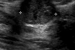
A 256 x 256-pixel ROI was manually centered for each mammogram at the microcalcification cluster and was positioned at the central breast region for each normal mammogram. The researchers compressed the images at 10:1, 25:1, 40:1, 70:1, and 100:1.
The resulting total set of 340 mammograms was then matched with the original versions. Over four reading sessions, each pair was presented in random order to the radiologists, who were asked to independently evaluate the quality of each compressed ROI compared to the original image using a five-point scale. A single rating score was given for each compressed ROI and encompassed the number and morphology of the microcalcification within the clusters as well as the mammographic parenchyma texture.
Each reading session was repeated after four weeks to assess inter- and intraobserver agreement.
Quantitative evaluation
The researchers then considered metric categories, including pixel-difference metrics for calculating the distortion between the compressed and original image on a pixel-wise basis; correlation-based metrics for quantifying the similarity between two digital images in terms of the correlation function; and a structural similarity (SSIM) index for quantifying structural changes.
The researchers found a strong correlation for the quantitative metrics of mean square error (r = 0.825), mean absolute error (r = 0.823), peak signal-to-noise ratio (r = -0.825), and structural similarity (r = -0.826) with the pooled radiologists' ratings. These metrics also produced the highest area under the ROC curve (0.922, 0.920, 0.922, and 0.922, respectively), according to the authors.
The researchers then used the highest accuracy values for each quantitative metric to define metric cutoff values, which were, in turn, employed to suggest a visually lossless compression ratio threshold.
This threshold was estimated to be between 25:1 and 40:1, "with 25:1 being a safe (a more conservative) [compression ratio] threshold above which the quality of compressed mammograms is significantly degraded," the authors wrote.