Spectral mammography used during breast screening can measure tissue density with high accuracy -- potentially improving the assessment of breast cancer risk, according to research presented at the American Association of Physicists in Medicine (AAPM) annual meeting in Charlotte, NC.
And in an era when the efficacy of mammography continues to be hotly debated, the technology could help doctors determine how often a woman should undergo mammography, or whether she needs further testing, according to researchers from the University of California, Irvine.
A woman with extremely dense breasts has up to four times the risk of breast cancer compared to a woman with fattier breasts, but standard mammography faces major challenges in accurately measuring breast density, said lead researcher Sabee Molloi, PhD, in a teleconference.
"Breast density is a major risk factor for cancer, and spectral mammography provides new information to help identify high-risk patients," he said. "Yet density is the most underexamined risk factor, due to a lack of standardization [in measuring it]."
Spectral mammography compared to standard mammography is like color television compared to black and white, Molloi said. Spectral mammography allows the image to be viewed at two different energy levels instead of just one, helping to quantify the density of a woman's breasts and, in turn, her relative risk.
Molloi and colleagues used Philips Healthcare's MicroDose photon-counting full-field digital mammography (FFDM) unit for the study, electronically upgrading it so that it provided spectral, or energy information. The team used the technology to image four models of breasts, representing different thicknesses ranging from 2 cm to 8.5 cm and densities ranging from 0% to 100%.
The results showed that spectral mammography could measure volumetric breast density in a screening exam with better than 2% accuracy, Molloi said. The technology also reduced the standard mammography radiation dose by 50%.
 |
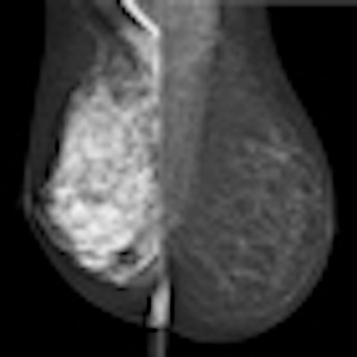
| Spectral mammography images: left is high-density breast tissue; right is low density. Image courtesy of AAPM |
Given that the MicroDose unit is available for clinical use in the U.S. and internationally, could the spectral mammography method Molloi's team developed be applied to every mammogram, providing an automatic breast density assessment? Yes, Molloi said.
"As it is now, the system needs to be upgraded [to include spectral mammography capability]," he said. "But when those upgrades are done, every mammogram acquired used for normal screening use could also provide breast density data."
Molloi and colleagues plan to further test spectral mammography in pilot studies of women as part of regular screening, he said.