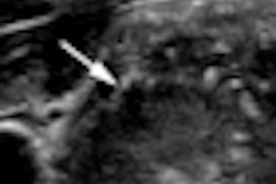
An elastography technique called supersonic shear imaging (SSI) aids breast lesion characterization when used with B-mode ultrasound, according to a new study from France published in the July issue of Radiology.
Researchers at the Institut Curie in Paris developed the technique, which uses a conventional ultrasound probe to perform two tasks: Rather than using mechanical external compression in a breast exam, the system generates mechanical vibration using acoustic radiation force created by a focused ultrasound beam; a fast ultrasound acquisition sequence (5,000 frames per second) captures the spread of shear, or transverse, waves (Radiology, July 2010, Volume 256:1, pp. 297-303).
"Our purpose was to determine the appearance of breast lesions at quantitative ultrasound elastography by using SSI and to assess the correlation between quantitative values of lesion stiffness and pathologic results," wrote lead author Alexandra Athanasiou, MD, and colleagues.
Displacement generated at the breast lesion creates a shear wave that conveys information about the properties of the tissue, providing a quantitative measure of elasticity, according the authors.
"With imaging in real-time, the propagation of the shear wave enables the recovery of the local shear wave speed and, consequently, the mapping of local tissue stiffness," they wrote.
Forty-six women received conventional ultrasound and SSI quantitative elastography between September 2006 and March 2007. There were 48 nonpalpable breast lesions found with B-mode ultrasound (28 benign, 20 malignant; mean size, 14.7 mm).
Although mammograms were available in all cases, the lesions weren't imaged well with mammography because of their small size or because the breast tissue was dense. Pathology results were available for all cases.
Quantitative lesion elasticity was measured in kilopascals (kPa). The team used an Aplio XG ultrasound unit (Toshiba Medical Systems Europe, Zoetermeer, the Netherlands); for the SSI sequences, it used a modified system (ATL HDI 1000, Philips Healthcare, Andover, MA).
Laboratoire Ondes et Acoustique, École Supérieure de Physique et de Chimie Industrielles, in Paris, holds the patent for the technique. SuperSonic Imagine, of Aix-en-Provence, provided the equipment and technical support for the study, according to the team.
The radiologist from the team performing the SSI first obtained saved B-mode images on the system hard drive, then did two SSI sequences each consisting of three successive pushing beams lasting three seconds. Total time for this exam was less than five minutes.
The vibration force was created by focusing ultrasound beams at five different depths; successive focusing depths were separated by 5 mm, making the pushing line 30-mm long, and each pushing focusing beam was a 150-µsec burst at 5 MHz. This supersonic energy made plane shear waves that spread transversally through the tissue in a few tens of milliseconds, Athanasiou and colleagues wrote.
The team found that SSI detected all breast lesions. Malignant lesions showed a mean elasticity value of 146.6 kPa ± 40.05, while benign lesions had an elasticity value of 45.3 kPa ± 41.1 (p < 0.001). These preliminary results indicate that not only does SSI help find breast masses and characterize cystic ones, it also could be easier to use, according to the researchers.
"From the user's perspective, one of the major differences of SSI compared with conventional elastography is that the mechanical vibration is induced automatically by the system by using the radiation force of the ultrasound beams," they wrote. "The reliability of the imaging technique does not depend on the skills of the sonologist in correctly vibrating or stressing the tissue."
By Kate Madden Yee
AuntMinnie.com staff writer
June 16, 2010
Related Reading
Breast elastography gets high sensitivity marks in 7-year study, November 11, 2009
Elastography shines in assessing breast lesions, April 29, 2009
Ultrasound elastography shows potential in thyroid nodules, February 11, 2009
Ultrasound elastography shows strength for diagnosing rotator cuff tears, January 15, 2009
Adding sonography to yearly mammograms helps find metachronous cancers, February 25, 2008
Copyright © 2010 AuntMinnie.com