Lower levels of carotid artery stenosis tend to worsen much more slowly than more significant stenosis, and they can be followed up with carotid ultrasound studies less frequently, according to researchers from the Ochsner Clinic Foundation in New Orleans.
In a retrospective analysis of nearly 3,000 patients over a 20-year period, the research team studied how various degrees of carotid artery stenosis tend to progress over time. They then developed follow-up recommendations based on the probability of progression for the patient's particular category of stenosis.
"We found that different degrees of carotid stenosis progress at different rates and, therefore, should be followed at different intervals," said Dr. Garrett Bennett.
He shared the findings during a presentation at the recent American Roentgen Ray Society (ARRS) annual meeting in San Diego.
Evolution of carotid disease
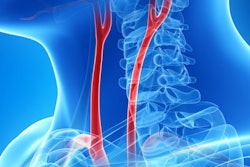
Carotid artery disease is a major cause of strokes and transient ischemic attacks, but not much is known about the disease's natural evolution. As a result, the Ochsner researchers set out to determine the rate of carotid stenosis progression over time, and then, based on these findings, to make recommendations for when interval follow-up should be performed with carotid duplex sonography exams. They also sought to determine if progression rates for carotid stenosis were influenced by plaque characteristics, patient symptoms, and laterality, Bennett said.
"We do not know of any good evidence-based recommendations currently for how to follow patients who either have carotid disease or are suspected of having carotid disease, particularly the lower degrees of stenosis," he said.
The team retrospectively collected 40,606 individual carotid artery exams from 16,478 patients over a 20-year period. All patients who received at least one carotid artery ultrasound exam were included in the study, while those who received carotid endarterectomy or carotid stenting were excluded. The degrees of carotid stenosis were classified according to a modification of nationally accepted criteria, and progression was defined as a change to a higher degree of stenosis from one examination to the next, according to the group.
Patients were classified as symptomatic or asymptomatic based on the indication of their initial study. In addition, the national criteria were used to determine if plaque morphology was heterogeneous or homogeneous. The researchers also compared the rates of stenosis and progression between the right and left carotid arteries.
Patients found to have 60% or greater stenosis are often closely followed in vascular surgery or cardiology laboratories for possible interventions, so the researchers elected to focus on carotid arteries with 1% to 39% stenosis and 40% to 59% stenosis. The team analyzed rates of carotid artery stenosis for 2,907 of these patients who received follow-up ultrasound exams.
The probability of carotid stenosis progression over 20 years was as follows:
- 1% to 39% stenosis: 7%
- 40% to 59% stenosis: 32%
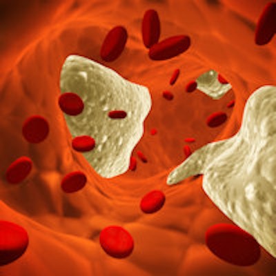
The researchers also found that symptomatic carotid stenosis patients had an 8% progression rate, while asymptomatic patients had a 9% progression rate. Only eight (3%) of the 261 vessels with heterogeneous plaque progressed, compared with 454 (7.8%) of the 5,814 vessels with homogeneous plaque. As for laterality, 304 (7.9% of follow-up exams) of right-sided exams showed progression, compared with 322 (8.7% of follow-up exams) of left-sided exams.
Follow-up recommendations
After analyzing how stenosis tends to progress, the researchers developed recommendations for when to perform follow-up examinations, broken down by patients with 1% to 39% carotid stenosis bilaterally, 40% to 59% stenosis bilaterally, or a combination of 1% to 39% and 40% to 59% stenosis for each carotid artery.
These recommendations were timed around the probability of a 10% increase in stenosis progression. The first follow-up scan should be performed when 10% of patients would have stenosis progression in at least one carotid artery; subsequent follow-up intervals would then be tied to each 10% increase in probability of having at least one progression in stenosis.
For example, 10% of patients who have both carotid arteries with 1% to 39% stenosis will have progression at three years, while seven years later 20% of these patients will have at least one progression in stenosis. By 10 years, 30% of patients will have at least one progression, and by 11.5 years, 40% of patients will have at least one progression, Bennett said.
Consequently, follow-up ultrasound studies should be performed at those time intervals and at subsequent times associated with a further 10% increase in the probability of stenosis progression.
| Recommended follow-up times based on degree of carotid artery stenosis | ||
| Both arteries with 1%-39% stenosis | One artery with 1%-39% and one with 40%-59% stenosis | Both arteries with 40%-59% stenosis |
|
3 years 7 years 10 years 11.5 years 13 years 14 years 15.5 years |
1 year 1.5 years 2.5 years 3.5 years 4.5 years 6.5 years 7.5 years 9 years 10 years 15 years |
0.5 years 1 year 1.5 years 2 years 3 years 4 years 4.5 years 6 years 8 years |
The researchers acknowledged a number of limitations of their study, including its retrospective nature and the unreliable reporting of patient symptoms by the ordering physicians in the electronic medical record. Because there was an extremely low incidence of heterogeneous plaque in their study, the researchers are also uncertain whether all plaques were appropriately categorized, Bennett said. They plan to investigate this issue in the future.
They also did not look into which patients received medical management in between examinations and how effective that was in preventing stenosis progression. In addition, they did not analyze the risk factors known to contribute to carotid disease, but they are currently reviewing this for a future study, according to Bennett. The study also did not include a cost-benefit analysis.
"Additional studies may be helpful in stratifying patients by both their clinical risk factors as well as their plaque characteristics and degrees of stenosis," he said.