Thanks to its ability to measure synovitis and detect joint damage, ultrasound may be a useful and less expensive tool to examine the effects of rheumatoid arthritis (RA), according to research presented at this week's American College of Rheumatology annual meeting in Washington, DC.
A research team led by Dr. Flávia Machado from the Universidade Federal de São Paulo in Brazil found that quantitative measures of RA-related joint findings were detected in nearly all of the joint recesses studied.
Machado and colleagues conducted the study to examine how quantitative and semiquantitative ultrasound measurements might indicate RA in small, medium, and large joints. They gathered quantitative measurements of synovial recesses and semiquantitative measurements of synovial hyperplasia, power Doppler, and bone erosions on an average of 6.348 joint recesses in 78 healthy volunteers and 60 patients diagnosed with RA.
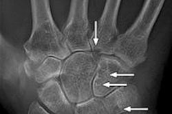
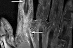
In addition to finding quantitative measures of RA in almost all joint recesses, semiquantitative measurements showed that greater scores in the radiocarpal and ulnocarpal joints of the wrist and the fifth metatarsophalangeal (MTP) joint of the foot increased the likelihood of RA, according to the American College of Rheumatology.
While in some cases analysis of ultrasound scores showed no statistical differences between healthy and RA-affected joints, this could be due to RA affecting each joint differently in individual patients, according to the researchers. In addition, some of the RA patients had few symptoms in some of the recesses studied, such as the hip and glenohumeral joints.
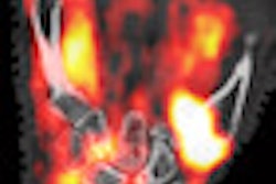
In general, however, measurement of RA patients showed greater scores of synovial hypertrophy and power Doppler capitation. Visualization of bone erosion from ultrasound also greatly increases suspicion of RA, according to the researchers.