SAN FRANCISCO - Sonography is proving to be a noninvasive and inexpensive way to measure tumor response to therapy, according to presenters at the Translational Research in Radiation Oncology and Radiology meeting, sponsored by the American Society for Therapeutic Radiology and Oncology (ASTRO).
Dr. Charles Cho and colleagues from Sunnybrook Health Sciences Centre, the University of Toronto, and Ryerson University, all in Toronto, discussed their animal research in a poster presentation at last week's meeting. They used high-frequency ultrasound spectroscopy to measure apoptosis in non-Hodgkin's lymphoma (NHL) cells after chemoradiation.
"NHL is expected to undergo apoptosis in response to both radiation therapy and chemotherapy.... The byproducts of apoptosis are thought to cause backscatter of high-frequency sound waves detected by ultrasound," they wrote.
The validity of sonography in this setting has been verified by one of Cho's co-authors, Dr. Gregory Czarnota, in previous research. "High-frequency ultrasound imaging devices operate at 30-50 MHz and offer increased resolution as well as the emerging capability to detect cells and tissues in different physiological states, including those undergoing programmed cell death, or apoptosis," Czarnota wrote in Methods in Molecular Biology (April 2002, Vol. 203, pp. 257-277).
 |
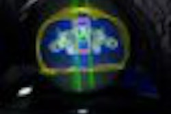
Above, ultrasound image of NHL xenograft done on a 20-MHz transducer at five hours after CHOP chemotherapy. The apoptotic areas correspond to the areas of high-intensity backscatter. Below, corresponding histology image with TUNEL stain. The areas that are stained brown correspond to the apoptotic areas. Images courtesy of Dr. Charles Cho.
 |
Most recently, Czarnota and colleagues integrated backscatter coefficient values correlated with the size of the nuclei in myeloid leukemia cells, human epithelial kidney cells, and breast cancer cells (Ultrasound in Medicine and Biology, March 2007, Vol. 33:3, pp. 389-401).
For the study led by Cho, NHL tumors were placed in severe combined immunodeficiency disease (SCID) mice. These tumor-bearing animals were then treated with single doses of 8 Gy or with one to two cycles of cyclophosphamide, hydroxydaunomycin, vincristine, and prednisone (CHOP) chemotherapy.
The animals were then imaged with 20-MHz spectroscopic ultrasound (VS40B, VisualSonics, Toronto). For the 26 mice that had chemotherapy, imaging was done several times, five to 72 hours after treatment. For the eight mice that had radiotherapy, sonography was performed once, six hours after treatment. Tumor sections were terminal transferase dUTP nick end labeling (TUNEL) stained to confirmed apoptotic cell death.
 |
Above, ultrasound image of NHL xenograft done on a 20-MHz transducer at 24 hours after CHOP chemotherapy. The apoptotic areas correspond to the areas of high-intensity backscatter. Below, corresponding histology image with TUNEL stain. The areas that are stained brown correspond to the apoptotic areas. Images courtesy of Dr. Charles Cho.
 |
The authors found a time-dependent increase in backscatter following CHOP chemotherapy, they wrote in their results. The measured high-intensity (10-decibel increase) patch areas on ultrasound were 1.76 mm² at five hours, 0.27 mm² at 12 hours, 2.03 mm² at 24 hours, 2.93 mm² at 48 hours, and 0.12 mm² at 72 hours.
Based on spectroscopy results, the midband fits changes from -51 dBr to -44 dBr after chemotherapy. The spectral slope was invariant at 0.66 dBr/MHz pretreatment and -0.60 dBr/MHz at 48 hours post-treatment.
"Image analysis demonstrated a correlation between the size of high-intensity patches on high-frequency ultrasound and immunohistochemical TUNEL staining or apoptotic areas," the group wrote.
 |
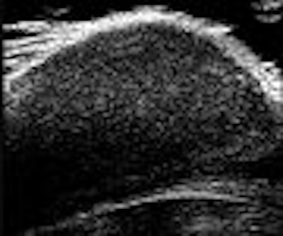
Above, ultrasound image of NHL xenograft taken after treatment with 8 Gy of radiotherapy. No areas of apoptosis or high-intensity backscatter were observed. Below, corresponding histology image with TUNEL stain. There are no areas of apoptosis seen on histology. Images courtesy of Dr. Charles Cho.
 |
However, the analysis of the ultrasound data after radiation treatment did not reflect the cell death seen on histology, they added. Cho told AuntMinnie.com that traditionally, NHL is very radiosensitive, but that the particular cell line that they used in the study may have been rendered insensitive. Also, during a discussion of Cho's poster, meeting program committee member Dr. Theodore Lawrence, Ph.D., pointed out that radiotherapy results may have differed if more than one time point had been measured post-treatment.
Lawrence also asked about the depth of the tumor visualized with high-frequency ultrasound. Cho acknowledged that with 40 MHz, the maximum depth generally ranges from 8 mm to 1 cm. While the 20 MHz used in this study did allow for greater imaging depth, it came at the cost of lower resolution, he said.
Cho said his group has run this same ultrasound-based apoptosis test for melanoma after photodynamic therapy and prostate cancer after radiotherapy, with similar results. Another area the team is currently looking at is locally advanced breast cancer to see if sonographically proven apoptosis can separate responders to neoadjuvant therapeutics versus those who are not responding, he said. Finally, they are working with contrast agents and microbubbles as well.
By Shalmali Pal
AuntMinnie.com staff writer
September 13, 2007
Related Reading
Blocking angiogenesis may render tumors radiosensitive, June 21, 2007
FDG uptake correlates with survival, biomarkers in NSCLC, June 19, 2007
Tetracycline analogues induce apoptosis in colon cancer cells, March 14, 2006
Copyright © 2007 AuntMinnie.com