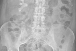
Digital photos of patients taken at the time of an imaging study can be easily integrated with the exams for viewing by radiologists on PACS, potentially decreasing errors and improving diagnostic capabilities at a very low cost, a group from Emory University has found.
In an article published online February 14 in the Journal of Digital Imaging, a team led by Dr. Senthil Ramamurthy discussed how an Android-based camera device and an integration server that communicates wirelessly with the institution's PACS network could be inexpensively deployed.
Digital patient photos could help reduce medical errors by decreasing the number of mislabeled exams, in addition to improving physician efficiency due to less time spent searching for anatomical landmarks. In many cases, patient photos could add to the diagnostic value of imaging studies and speed up interpretation, according to the researchers.
"Implementation of our technique for simultaneous acquisition and display of photographic and medical imaging data is feasible with existing technologies," the authors wrote. "Thus, the technology is available for integration of digital cameras for acquisition of patient facial photographs at the point of care of medical imaging for a wide variety of modalities."
Preliminary experience with a prototype device mounted on a portable x-ray system was presented by senior author Dr. Srini Tridandapani, PhD, at the 2012 Society for Imaging Informatics in Medicine (SIIM) meeting, but the researchers shared more details and analysis of the system in the current article.
"These digital photographs will be small additions to the imaging study similar to the scout or localizer images that are performed with CT studies," the authors wrote. "We do not intend these digital photographs to entirely replace numerical identifiers, but rather we envision that they would supplement and strengthen these identifiers. However, in some cases, such as unconscious trauma patients, these photographs may indeed be the only available identifiers."
Off the shelf
The group custom-built the camera device using off-the-shelf components, assembled around a BeagleBoard-xM platform with an ARM microprocessor. It includes a 3-megapixel camera, a Bluetooth-connected radiofrequency identifier (RFID) reader, and an 802.11 b/g wireless module for connecting to the integration server.
Mounted on a portable computed radiography system, the prototype camera device operates transparently to the technologist. When the technologist presses the x-ray system's trigger, it also automatically activates the camera device. The photo is taken and the RFID reader scans the cassette ID.
The integration server retrieves current studies from the PACS, reads the cassette ID from the DICOM header, and matches the patient photo based on the cassette ID and time stamp. A new series is created with the matched, DICOM-converted photo and sent to the PACS, according to the authors. A hanging protocol then displays the patient photo along with the images.
In addition to its utility for digital radiography and portable x-ray, the device could also be adapted for use with ultrasound, CT, MRI, and PET, the researchers noted.
In an observer study of their system conducted at Emory, radiologists were only able to detect 12.5% of misidentification errors in radiograph pairs without patient photographs; however, they were able to detect 64% of errors in radiograph pairs with patient photos.
That study will be published in the April issue of the American Journal of Roentgenology. Results from an additional observer study involving 87 radiologists from around the U.S. are currently being submitted for publication, Tridandapani told AuntMinnie.com.
Cost analysis
Because a 3-megapixel camera can provide a JPEG-compressed picture for less than 0.5 MB, the additional overhead for adding a single photo to a medical imaging study would range from 12.5% for a one-view chest radiograph to 0.05% for a heart CT exam. One megabyte of memory costs less than 1 cent, so the added cost is negligible relative to the cost of the examination, according to the researchers.
The team expects that "snap-on" kits to retrofit existing imaging devices could be developed for $200 or less per kit. The authors noted that approximately 137 imaging devices are in use at Emory's affiliated hospitals and clinics; retrofitting all of the machines would cost less than $30,000.
"While it is difficult to estimate the extent of the impact that our proposal will have on patient safety, it should not be difficult to argue that $30,000 U.S. would be a fairly inexpensive investment if even one major complication or death were to be prevented out of 480,000 medical imaging studies performed at our institution annually," the authors wrote.
The Emory group is now working on improving the prototype for portable x-ray machines, exploring further miniaturization of the device, Tridandapani said.


