At least one gadolinium-based contrast agent (GBCA) appears to be steering clear of problems related to gadolinium retention. Researchers in South Korea found no evidence of gadolinium retention following multiple administrations of the agent gadoterate meglumine, according to a study published online in PLOS One.
The study of 385 patients found no significant difference in dentate nucleus-to-pons and globus pallidus-to-thalamus signal-intensity ratios between unenhanced T1-weighted images and subsequent MRI scans that used gadoterate meglumine (Dotarem, Guerbet) as a contrast material. The results were the same even for a subset of 33 patients who received 20 or more administrations of the macrocyclic GBCA (PLOS One, August 31, 2017).
"As the dentate nucleus is the location where most gadolinium accumulation occurs, the lack of any observable T1 signal changes in the dentate nucleus may support the current clinical use and recommendation of a macrocyclic GBCA in guidelines with safety concerns," wrote co-lead authors Dr. Ji Ye Lee and Dr. Ji Eun Park and colleagues from Soonchunhyang University Bucheon Hospital and Asan Medical Center.
Gadolinium factor
Over the past few years, several studies have revealed that minute traces of gadolinium can remain in certain regions of the brain long after GBCA injection, manifesting as greater T1 signal intensity during unenhanced MRI scans. In comparisons of various GBCA types, researchers have found that macrocyclic agents have reduced signal intensity -- or none at all -- compared to linear agents.
For example, a March 2017 study by Radbruch et al found no evidence of increased signal intensity in the dentate nucleus of pediatric patients who had received gadoterate meglumine, a macrocyclic ionic GBCA. The same researchers came to a similar conclusion in a December 2016 study that evaluated signal intensity from gadoterate meglumine and another macrocyclic GBCA, gadobutrol (Gadavist, Bayer HealthCare).
While other clinical studies have reported a change in T1 signal intensity for the three commercially available macrocyclic GBCAs -- gadoteridol (ProHance, Bracco Diagnostics), gadobutrol, and gadoterate meglumine -- Lee, Park, and colleagues noted that the increases were not statistically significant.
Gadoterate meglumine has been used as the primary GBCA at their institution since 2009, according to the researchers. To explore any potential adverse effects from the agent, they investigated various clinical situations, including patients who received more than 20 administrations, as well as the time interval between MRI scans and whether patients received chemotherapy or radiation therapy.
Retrospective review
The group reviewed records dating from March 2009 through July 2016 and found a total of 385 patients who met the study's inclusion criteria. The 213 women and 172 men had a mean age of 56.8 years (range, 18-90 years), and none presented with severely impaired renal function or acute renal failure. Approximately half (52%) of the patients were referred for the evaluation of metastases.
All scans were performed on a 1.5-tesla (Achieva, Philips Healthcare) or 3-tesla MRI scanner (Achieva or Ingenia, Philips, or Skyra, Siemens Healthineers) with an eight-channel head coil. Patients were given a standardized single dose of 13 mL of gadoterate meglumine.
Regions of interest (ROIs) targeted the right dentate nucleus, central pons, right globus pallidus, and right thalamus. Two readers then compared the signal intensities of the four brain regions for the first unenhanced MRI scan and the last GBCA-enhanced scan. To determine any differences, signal-intensity ratios were calculated for the dentate nucleus and pons and for the globus pallidus and thalamus.
| Signal-intensity ratios in patients with normal renal function | ||||
| Mean signal-intensity ratio | Unenhanced MRI | Gadoterate meglumine-enhanced MRI | Ratio difference | p-value |
| Dentate nucleus-pons | 1.021 ± 0.05 | 1.021 ± 0.06 | 0.001 ± 0.063 | 0.697 |
| Globus pallidus-thalamus | 1.029 ± 0.08 | 1.006 ± 0.06 | -0.021 ± 0.083 | < 0.001 |
Only patients with abnormal renal function showed a significant increase in the mean dentate nucleus-pons signal-intensity ratio difference (0.018 ± 0.038, p = 0.019), which differed significantly from patients with normal renal function (p = 0.019).
Interestingly, the mean globus pallidus-thalamus signal-intensity ratio showed a significant decrease from the first unenhanced MRI scan (1.029 ± 0.08) to the last GBCA-enhanced scan (1.006 ± 0.06) (p < 0.001). This downward trend was significantly correlated to patients with six or more GBCA administrations (p = 0.01), mean time of fewer than 90 days between scans (p = 0.001), and number of radiation therapy sessions (p = 0.022).
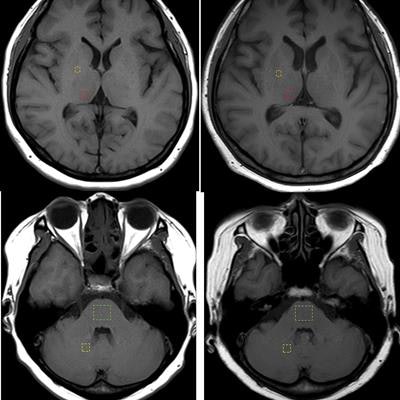
 Axial T1-weighted MR images show regions of interest in the quantification of signal intensity (dashed lines) for the globus pallidus, thalamus, dentate nucleus, and pons. Globus pallidus and thalamus images (upper row) are from the first scan and after 21 administrations of macrocyclic ionic GBCA. Also shown are the dentate nucleus and pons (lower row) from the first scan and after 21 administrations of macrocyclic ionic GBCA. Image courtesy of PLOS One.
"Our research supports the current extensive use of macrocyclic GBCAs, particularly the use of macrocyclic ionic agents," the researchers concluded. "Future studies using large clinical data to compare different macrocyclic GBCAs may be helpful to assess their in vivo stability and safety."
The group cited several limitations to the study, including its retrospective approach. In addition, only four brain regions were selected for sources for signal intensity.
"It is impossible to completely rule out intracranial gadolinium deposition in other sites," the researchers wrote. "However, there are no alternatives since the dentate nucleus and globus pallidus are recognized as the sites with the highest in vivo accumulation of gadolinium."