Patients who received more than 35 injections of gadolinium-based contrast agents (GBCAs) had higher levels of signal intensity on unenhanced MR images of the brain, according to a new study published online August 11 in Radiology. But researchers weren't able to find any adverse clinical effects in the individuals.
The group from Weill Cornell Medicine and Columbia University Medical Center found greater signal intensity in the dentate nucleus and global pallidus in patients who had a large number of gadolinium administrations, compared with individuals who had fewer GBCA administrations. Despite the rise in signal intensity, there were no obvious signs that the additional gadolinium caused any harm to patients.
"The most important point is that in spite of patients receiving more than 35 administrations of GBCA with subtle signal changes in several brain nuclei, we could not identify any adverse clinical effects related to this," study co-author Dr. Martin Prince, PhD, professor of radiology at Weill Cornell Medicine, told AuntMinnie.com.
GBCA ramifications
The presence of minute gadolinium traces in the brain years after MRI contrast administration has been well-documented in the past few years. In the current paper, Prince and colleagues cited the study by Kanda et al that found high signal intensity on unenhanced T1-weighted MRI scans in the dentate nucleus and globus pallidus of the brain in 19 patients who had received six or more GBCA injections.
After reviewing the records of one patient who received 34 GBCA administrations at their institution and noticing increased signal intensity beyond the dentate nucleus and globus pallidus, Prince and colleagues pondered the ramifications of a large number of GBCA injections for patients.
"The huge issue is: Is it of any importance, because no one has been able to identify any clinical sequelae?" he said. "Are we seeing the effects of those very trace amounts, which is of negligible consequence, but exciting because we are seeing it on [MR] images? Or is there real clinical importance and something we should avoid?"
The researchers reviewed patient information and MR images at both facilities from between January 2000 and August 2015 to find subjects who met the study's criteria. Among other stipulations, the individuals had to have received 35 or more administrations of a linear GBCA, which at their facility included gadodiamide (Omniscan, GE Healthcare), gadopentetate dimeglumine (Magnevist, Bayer HealthCare), or gadobenate dimeglumine (MultiHance, Bracco Diagnostics).
Patients who received macrocyclic agents were excluded. Also, the subjects had to have a baseline initial T1-weighted brain MRI exam performed before the first GBCA administration; those without baseline images were excluded.
In all, 13 subjects met the study criteria. The mean number of GBCA administrations was 43; the total numbers of administrations ranged from 39 to 59 (Radiology, August 11, 2016).
Initial brain MR imaging was performed primarily on 1.5-tesla scanners, with later exams conducted mostly on 3-tesla devices (GE, Siemens Healthineers, Philips Healthcare). The final analysis included 550 studies, of which 72 scans were from outside institutions.
To detect signs of residual gadolinium enhancement, three radiologists independently interpreted unenhanced T1-weighted MR brain images after six, 12, and 24 GBCA administrations, and after the last contrast injection. Their task was to identify any areas of increased T1 signal intensity, compared with the baseline MRI results, and measure the rate of signal increase per injection.
GBCA hot spots
All three radiologists found increased signal intensity in the dentate nucleus, globus pallidus, and substantia nigra in all 13 subjects, as well as greater signal intensity in regions such as the posterior thalamus, red nucleus, and colliculi in other subjects. The dentate nucleus also had the greatest mean increase in signal intensity, nearly double that of the globus pallidus and posterior thalamus.
| Regions of increased T1 signal intensity | ||
| No. of patients | Mean increase per administration | |
| Dentate nucleus | 13 | 0.53% |
| Globus pallidus | 13 | 0.23% |
| Substantia nigra | 13 | 0.25% |
| Posterior thalamus | 12 | 0.26% |
| Red nucleus | 10 | 0.25% |
| Colliculi | 10 | 0.26% |
| Cerebellar peduncle | 7 | 0.19% |
| Head of caudate nucleus | 4 | 0.02% |
| Body of caudate nucleus | 4 | 0.14% |
| Putamen | 2 | -0.01% |
The increase in signal intensity in the dentate nucleus was much more dramatic than what Kanda et al found in the same area, Prince and colleagues noted. The current research also differed from previous studies in discovering greater signal intensity in other brain regions.
"When there are more than 35 gadolinium injections, we are seeing more locations in the brain that show signal change beyond the dentate nucleus and global pallidus," Prince said.
But while they found signs of gadolinium deposition, the phenomenon appeared to have no clinical effect on patients.
"We looked and looked and could not find any pattern of clinical impact," he said. "It continues to be a topic of mystery and interest and of uncertain impact on the practice of radiology."
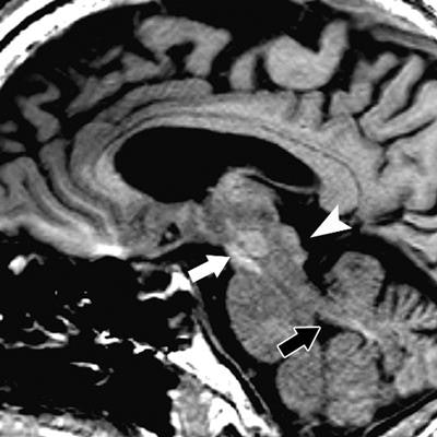
 T1-weighted MRI of a 61-year-old man with left frontal glioblastoma shows high signal intensity in the colliculi (arrowhead), red nucleus and substantia nigra (white arrow), and superior cerebellar peduncle (black arrow) after 35 GBCA administrations, compared with baseline MRI prior to GBCA. Images courtesy of Radiology.
As research continues into the long-term effects of GBCAs, Prince emphasized there have been no changes in the utilization of gadolinium-based contrast agents at Weill Cornell Medicine and Columbia University Medical Center.
"We are continuing to see year-over-year growth in the clinical demand for MRI, including MRI with gadolinium, in order to solve clinical issues to help with patient care," he added.
As for future plans, the researchers remain very interested in investigating the clinical effects of multiple GBCA administrations on patients undergoing MRI scans of the brain and other parts of the body.
"Even if there are no clinical sequelae, there is still the issue that the brain looks a bit different on unenhanced T1-weighted images," he said. "If we see a bright signal in these hot spots, that may be a sign of previous gadolinium injections rather than a sign of pathology. That is important to know."