Researchers at Massachusetts General Hospital (MGH) have developed a method that combines diffusion-weighted MRI (DWI-MRI) and key clinical characteristics to determine which severe stroke patients would most likely benefit from catheter-delivered removal of blood clots.
The study, published online November 6 in JAMA Neurology, showed that use of the technique enabled MGH physicians to screen stroke patients and determine who should be sent on for interventional blood-clot removal: More than half the patients deemed suitable for the technique had recovered well three months later. Meanwhile, fewer patients who were judged as unsuitable recovered, as did patients who were evaluated with CT.
"Our work shows that MRI identifies patients with greater precision, which means that fewer patients are inaccurately excluded from treatment that might otherwise benefit them," commented lead author Dr. Thabele Leslie-Mazwi. "As endovascular therapy expands at a regional, national, and international level, we think this precision will be essential to providing the most personalized and cost-effective care."
Time is brain
The most critical task in treating ischemic strokes is to restore blood flow as quickly as possible to reduce the amount of brain tissue that may be destroyed, the authors noted. Mechanical thrombectomy with clot-removal devices delivered via catheter has been shown to be effective, but emergency physicians still have to decide which patients should get the treatment.
While many previous studies advocate the use of CT to achieve the best outcomes for patients who may be suitable for intervention, MGH primarily uses DWI in its clinical setting to "produce the most precise estimate of the size of the ischemic core -- the area of brain tissue that has been destroyed by the stroke," the authors wrote.
"The critical question has been how to optimize its use to benefit the most patients while minimizing any harm," added senior author Dr. Gilberto González, PhD, director of neuroradiology in MGH's department of radiology. "It appears that the MGH approach to patient selection is superior to other approaches and may be the optimal approach."
Who might benefit?
The MGH criteria separate stroke patients into those would be probably benefit from mechanical clot removal (a "likely-to-benefit" group) and those who wouldn't (an "uncertain-to-benefit" group).
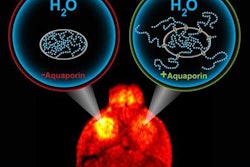
Figuring prominently in the criteria is DWI, which measures the movement of water molecules in vivo and indirectly reflects tissue microstructural characteristics. The researchers used DWI to measure the volume of infarcts; a recent Cleveland Clinic study found that patients with infarcts smaller than 70 mL might benefit from endovascular treatment.
Another component of the criteria is the modified Rankin Scale (mRS), used to characterize the degree of a person's disability after a stroke. The seven-point scale ranges from 0, which indicates no symptoms, to 6, which means the person has died.
The criteria used to determine which patients should receive mechanical clot removal are summarized below.
Likely to benefit:
- Younger than 80 years
- Less than six hours from stroke onset or last seen well to groin puncture
- Premorbid baseline mRS score of 1 or less
- Life expectancy of more than 12 months
- DWI volume less than 70 mL
Uncertain to benefit:
- 80 years of age or older
- Six to eight hours from stroke onset or last seen well to groin puncture
- Premorbid baseline mRS score of 2 to 3
- Life expectancy of more than six months
- DWI volume of 70 mL to 100 mL
To test the criteria, the researchers enrolled 103 consecutive patients who presented for acute stroke in a prospective study between January 2012 and December 2014. Both CT and CT angiography were performed on multidetector-row scanners (GE Healthcare) from the vertex to the aortic arch after injection of 65 mL to 140 mL of a nonionic contrast agent (Isovue, Bracco Diagnostics). The MRI protocol included DWI for all eligible patients with fluid-attenuated inversion-recovery (FLAIR) and gradient echo sequences.
Some subjects were scanned exclusively on CT due to pacemakers, metallic implants, and other contraindications for MRI. In some cases, CT findings indicated no evidence of an infarct or other abnormality from the stroke, so no MRI scan was necessary.
Seventy-two people received MRI scans, of whom 40 met the criteria for being likely to benefit from mechanical blood clot removal. Meanwhile, 32 subjects were classified into the uncertain-to-benefit group. The remaining 31 patients with similar ischemic strokes underwent endovascular treatment after a CT scan only.
Favorable outcomes
Primary outcomes were based on mRS scores after three months through a physician telephone interview or direct neurological clinical evaluation.
Of the 40 patients in the likely-to-benefit group, 21 (52%) had favorable outcomes; that is, complete functional independence. Of the 32 patients in the uncertain-to-benefit group, only eight (25%) had favorable outcomes. Among those who had only CT scans, there were nine (29%) favorable outcomes.
"Thus, the overall rate of favorable outcomes was 36.9% when treating one of every three patients screened," the authors wrote. "This may be close to the optimal trade-off with current treatment technology."
| Endovascular stroke treatment outcomes | |||
| Likely to benefit | Uncertain to benefit | CT only | |
| No. of patients | 40 | 32 | 31 |
| Favorable outcome | 21 (52%) | 8 (25%) | 9 (29%) |
| Successful reperfusion | 27 (67%) | 24 (75%) | 20 (64%) |
| Favorable outcome after successful reperfusion | 20/27 (67%) | 8/24 (75%) | 8/20 (40%) |
| Favorable outcome after failed reperfusion | 1/13 (8%) | 0/8 (0%) | 1/11 (9%) |
| Mortality | 7/40 (17%) | 8/32 (25%) | 9/31 (26%) |
While the positive results in the likely-to-benefit group are similar to findings in recent clinical trials, the key difference is that DWI resulted in a significantly greater number of screened patients receiving endovascular treatment, the researchers noted.
Thus, with the MGH criteria, they were better able to determine which patients would benefit from intervention.
"The next frontier to investigate is treatment six to 24 hours after stroke onset," said study co-author Dr. Joshua Hirsch, director of endovascular neuroradiology, in an MGH news release. "Data we have accumulated over the past decade suggests that many patients may be successfully treated at this late stage, and MRI is the most powerful means to accurately identify these individuals."
If that finding holds true, there may be time to transfer some patients who are initially seen at regional hospitals to facilities such as MGH, which may be better suited to conduct this type of screening program, he said.