Using MRI, Australian researchers found smaller brain size and other abnormalities that could lead to long-term developmental issues in babies born 32 to 36 weeks into gestation, according to a study published online June 10 in Radiology.
These moderate and late preterm babies account for approximately 80% of all preterm births and are responsible for much of the rise preterm birth rates over the past 20 years. However, there have been no large-scale studies published on brain alterations in this group that could provide insight into brain-behavior relationships.
Injury due to bleeding in the brain or a lack of blood flow, oxygen, or nutrition could explain some of the abnormal brain development that occurs in very preterm babies, said lead author Dr. Jennifer Walsh from the Royal Women's Hospital in Melbourne in a statement from RSNA. But in some preterm babies there may also be no obvious explanation for what appears to be slower brain development, she added.
Walsh and colleagues performed MRI exams on 199 moderate and late preterm infants and 50 infants born between 38 and 44 weeks of gestation (control group), looking for signs of brain injury. They also compared the size and maturation of multiple brain structures between the two groups (Radiology, June 10, 2014).
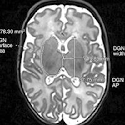
 Axial T2-weighted MR image (left) shows deep gray nuclei width, anteroposterior distance, and surface area. Coronal T2-weighted MR image (right) shows lateral ventricle atrial measures and transverse cerebellar diameter. Images courtesy of RSNA.
Injury rates were similar between the two cohorts; however, moderate and late preterm birth was associated with smaller brain size at term-equivalent age, the researchers found. In addition, moderate and late preterm infants had less developed myelination in one part of the brain and more immature gyral folding than the control group.
Myelination involves the formation of a fatty sheath around nerve fibers, while gyral folding is the process in which the cerebral cortex folds to increase brain surface area. Both are important parts of early brain development.
The findings suggest that moderate and late preterm birth could disrupt the trajectory of brain growth that normally occurs in the last two or so months in utero. "Given that brain growth is very rapid in the last one-third of pregnancy, it is perhaps not surprising that being born during this potentially vulnerable period may disrupt brain development," Walsh said.
Medication and early intervention have been effective in helping very preterm babies catch up to their peers. However, whether the existing treatments will help babies born between 32 and 36 weeks is unknown due to a lack of research, she said.
By learning more about brain function in this group, Walsh and colleagues hope to be able to develop different treatments to improve function and long-term outcomes.
The group also plans to follow the infants in the current study through childhood to learn more about the relationship between brain abnormalities and the infants' development. They are also assessing additional MRI information about brain structure and function in these children.