A novel MR imaging technique known as restriction spectrum imaging (RSI) can accurately differentiate aggressive prostate cancer from low-grade or benign tumors, according to a study published in the June 1 issue of Clinical Cancer Research.
Researchers from the University of California, San Diego (UCSD) and the University of California, Los Angeles (UCLA) found that RSI can also help guide biopsies and subsequent treatment (Clin Cancer Res, June 1, 2016, Vol. 22:11, pp. 2668-2674).
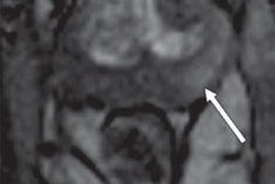
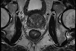
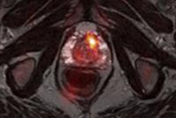
RSI-MRI corrects for magnetic field distortions found in other imaging techniques and focuses on water diffusion within tumor cells that exhibit a high nuclear volume fraction. This enables RSI to more accurately plot a tumor's location and allows for differentiation between tumor grades.
Senior author Dr. David Karow, PhD, an assistant professor of radiology at UCSD School of Medicine, said RSI allows clinicians to predict the grade of a tumor sometimes without a biopsy of the prostate tissue, while adding only 2.5 to five minutes to a pelvic MR scan in the study.
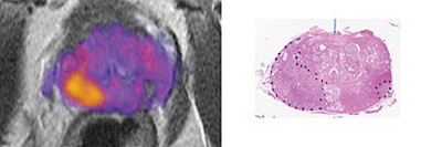
 MRI (left) shows the prostate enhanced with RSI. The higher-grade tumor is indicated in orange and yellow. The image on the right shows a digitized section of the prostate with the tumors outlined with dotted lines. Images courtesy of Clinical Cancer Research.
Greater accuracy helps customize treatment for each patient by identifying more aggressive cancers, which require immediate attention. The findings also suggest that RSI-MRI could eventually serve as a standalone, noncontrast screening tool that would take 15 minutes, compared with 40 to 60 minutes for a contrast-enhanced MRI scan.