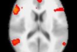
Functional MR images (fMRI) have shown the area of the brain that helps recognize facial expressions, according to a study published online April 19 in the Journal of Neuroscience.
Researchers from Ohio State University found that the posterior superior temporal sulcus became activated when subjects looked at images of people making different facial expressions; the group then took note of movement in specific parts of the face.
The researchers created a software algorithm that cross-referenced fMR images with the different facial muscle movements shown in the test photographs. They were then able to create a map of regions within the posterior superior temporal sulcus that activated for different facial muscle groups, such as the eyebrows or lips.
The algorithm achieved about a 60% success rate in decoding human facial expressions, regardless of the facial expression or the person viewing it.