Using SPECT/MRI to image sentinel lymph nodes to assess whether metastases are present could greatly benefit cervical cancer patients without enlarged lymph nodes, according to a study published in the April issue of the Journal of Nuclear Medicine.
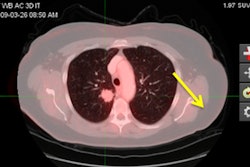
Researchers at University Medical Center Utrecht in the Netherlands used technetium-99m (Tc-99m) nanocolloid SPECT/MRI fusion to assess patients with early-stage cervical cancer as to whether their cancer had spread to the sentinel lymph nodes (JNM, April 2016, Vol. 57:4, pp. 551-556).
After excluding patients with enlarged lymph nodes as detected on MRI, the remaining cohort underwent a sentinel lymph node mapping procedure with preoperative Tc-99m nanocolloid SPECT/CT. By creating fused datasets of the SPECT and MRI, sentinel lymph nodes could be identified on MRI and accurately correlated with the histological result of each individual sentinel lymph node.
In 75 cases, 136 sentinel lymph nodes were analyzed; 13 nodes (10%) among eight patients contained metastases.
The authors noted that the key element of the study is the focused review of fewer than five sentinel nodes, instead of indiscriminately reviewing the entire pelvic lymphatic chain, which could contain as many as 100 nodes per patient.
The ultimate goal, they added, is to reduce morbidity through more informed decisions on whether to use radical hysterectomy or chemo/radiation therapy for each patient, rather than using only stage-based treatment selections.