MRI can be used to visualize inflammation in the pancreas that leads to type 1 diabetes, according to a study in the February 17 issue of the Proceedings of the National Academy of Sciences.
The findings from Joslin Diabetes Center and Massachusetts General Hospital could benefit research investigating how to slow down or stop type 1 diabetes at an early stage, and the study could also provide information on how diabetes progresses.
The researchers investigated the possibility of using MRI and ferumoxytol, a coated iron nanoparticle used as an iron replacement therapy, to image inflammation in the pancreas. Ferumoxytol leaks from blood vessels in areas of inflammation and is taken up by immune cells called macrophages, which accumulate at sites of inflammation.
Co-lead authors Dr. Jason Gaglia and Dr. Mukesh Harisinghani and colleagues recruited 11 patients with newly diagnosed type 1 diabetes and evidence of antibodies against the pancreas. They also included 10 controls with no sign or family history of diabetes (PNAS, February 17, 2015, Vol. 112:7, pp. 2139-2144).
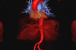
Ferumoxytol-MRI of the patient group showed clear evidence of ferumoxytol accumulation in the pancreas, according to the researchers, indicating ongoing inflammation. MR images from the control group did not show the same accumulation.
The MRI technique could help better define which patients will progress to diabetes and classify subgroups of patients who might benefit from different therapeutic strategies, the researchers speculated.