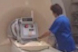
A team of researchers from Stanford University has developed dedicated pediatric MRI coil technology that enables the acquisition of scans in less time, potentially reducing the amount of anesthesia that needs to be used.
A group led by Dr. Shreyas Vasanawala, PhD, worked with engineers from GE Healthcare to develop parallel arrays of abdominal MRI receiver coils. The smaller coil size improves image quality, while the parallel array improves scan times by enabling individual coils to acquire signals simultaneously rather than sequentially.
The researchers have also applied an image processing technique called compressed sensing, a form of data undersampling that reduces scan times by gathering only a small fraction of the data that are usually needed to enable reconstruction of an image. Vasanawala and Michael Lustig, PhD, an engineer from the University of California, Berkeley, developed an algorithm that reconstructs the full MR image from a small number of data points in high fidelity. The team also applies a motion-correction algorithm to maintain clear images even if a child is breathing.
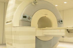
While all of these approaches have been researched in other areas of MRI, Vasanawala believes his team is the first to apply all the technologies to pediatric imaging. They report that at Stanford, pediatric scans that previously took an hour to acquire can now be performed in 10 minutes, and Stanford is now using less anesthesia than it did in the past.
Their approach is detailed in an article in the July 2014 issue of Magnetic Resonance in Medicine.