Volumetric functional MRI (fMRI) can reveal within a month whether chemotherapy is successfully killing tumor cells, long before liver or pancreatic tumors shrink or fail to shrink, according to two studies published in the July issue of Radiology.
Researchers from Johns Hopkins Medical Institutions believe this is the first time the diagnostic imaging procedure has been shown to add survival time by determining which patients might benefit from repeat or higher-dose chemotherapy.
The fMRI scans take only minutes to perform and can detect the movement of water molecules inside tumor cells, while specially developed software conducts the mathematical analysis to calculate the imaged tumor cells' apparent diffusion coefficient (ADC). The software (MR Oncotreat) used in the scans was developed at Johns Hopkins and at Siemens Healthcare, which also supplied the 1.5-tesla whole-body MRI scanner (Magnetom Avanto).
Living tumor cells show a low coefficient, as water absorption in and out of the cells is controlled, while dying cells have a relatively high coefficient, as the tumor cells' membranes lose the ability to restrict water movement, according to the authors (Radiology, July 2012, Vol. 264:1, pp. 97-109, 285-294).
Volumetric fMRI scans could help people with aggressive cancers learn quickly whether their treatment is working or they need to consider other treatment options, instead of waiting for months to see tumor shrinkage, said study senior investigator Dr. Ihab Kamel, PhD, associate professor at Johns Hopkins University School of Medicine, in a statement about the studies.
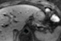
Results from one study showed that the high-precision 3D images, taken shortly before and one month after a form of chemotherapy known as transcatheter arterial chemoembolization, could assess treatment efficacy for cholangiocarcinoma, a rare and advanced form of liver bile duct cancer.
The study included 29 men and women, ages 29 to 82 years, who received treatment at the Johns Hopkins Hospital. The researchers found that 22 patients whose fMRI scans showed increases in ADC scores of more than 45% lived at least 10 months longer, while 17 patients with ADC score increases of more than 60% lived at least 17 months longer.
The other study, also at Johns Hopkins, tested the scans on 26 men and women, ages 37 to 79 years, who were diagnosed with islet cell carcinoma, a form of pancreatic cancer. The researchers analyzed 215 lesions from the 26 patients, scanning shortly before and one month after transcatheter arterial chemoembolization.
For the 78 tumors that responded well to chemotherapy, fMRI showed ADC score increases averaging at least 70%. In contrast, for the 137 tumors that did not respond, ADC score increases averaged less than 40%. Treatment was considered successful if tumor shrinkage continued for at least six months.
The ability to determine which tumors are responding to treatment is very important in islet cell carcinoma, Kamel said. Some symptoms of the disease -- such as headaches, ulcers, pain, and diarrhea -- can be masked by liver hormone production; the disappearance or absence of symptoms is not a reliable indicator of treatment success.
Additional research will include larger studies to measure how well ADCs for each kind of chemotherapy combination predict survival and how much time remains to switch treatment plans.
|
Study disclosures Siemens Healthcare provided funding support for both studies. Bayer HealthCare and Bracco Diagnostics also provided research grants. One study author is also supported by Biocompatibles, Genentech, Bayer HealthCare, Philips Medical, Nordion, Context Vision, CeloNova, and a 2000 RSNA Education Seed Grant. |