British researchers achieved 100% positive and negative predictive values using 7-tesla MRI and a specialized scanning protocol to scan the brains of patients suspected of having multiple sclerosis (MS). The technique could possibly be applied to 3-tesla magnets, providing a means to diagnose MS patients without further invasive workup.
Researchers from Nottingham University Hospitals NHS Trust in the U.K. believe their findings could lead to a single diagnostic test for the cause of MS, helping patients avoid additional, unnecessary procedures and expediting their diagnosis.
The findings were presented at the recent American Academy of Neurology (AAN) annual meeting by Dr. Niraj Mistry, a research fellow in the division of clinical neurology at the University of Nottingham.
An unmet need
"There is still an unmet need in multiple sclerosis diagnostics," Mistry told AuntMinnie.com. "Some patients currently suspected of having MS can wait a long time for diagnosis to be confirmed, often despite multiple tests, such as hospital MRI and spinal taps, and evoked potential testing."
White-matter lesions are frequently detected on brain MRI scans performed for a variety of indications, the researchers noted. Most lesions are caused by small-vessel ischemic disease and other conditions that can mimic multiple sclerosis on MRI. Most MS lesions are located around a vein, and this arrangement distinguishes MS from other causes of brain lesions.
For this clinical application, 7-tesla MRI has two main advantages.
"First, the improved signal-to-noise ratio allows higher-resolution images to be obtained, making visualization of smaller structures more reliable, including the internal structure of MS lesions," Mistry said. "Second, 7-tesla MRI allows us to better exploit local magnetic effects in brain tissues."
Specifically, deoxygenated blood in veins has different magnetic properties than oxygenated blood in arteries. Using a 7-tesla MRI technique known as T2*-weighted imaging, researchers can make veins appear especially dark, while MS lesions look bright. "Previously, it has not been possible to see this in live patients using a single brain scan," Mistry added. "Now, with T2* MRI, we can."
For this study, 25 patients were recruited from the neurology outpatient department at Nottingham University Hospitals NHS Trust. The subjects exhibited brain abnormalities on MRI that raised the possibility of demyelination, a degenerative condition that erodes the myelin sheath in the brain that protects nerve fibers. A diagnosis for the 25 patients could not be made through initial clinical and/or radiological assessment.
MR images were acquired on a 7-tesla scanner (Achieva, Philips Healthcare) and readers were blinded to the subjects' clinical data. Patients were followed by their neurologist for an average of 1.7 years.
Lesion location
While MS lesions are known to occur in all parts of the brain and spinal cord, the researchers focused their study on characterizing the white-matter lesions that are most easily detected using conventional MRI.
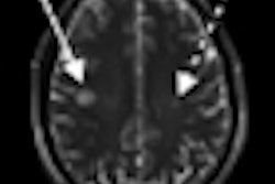
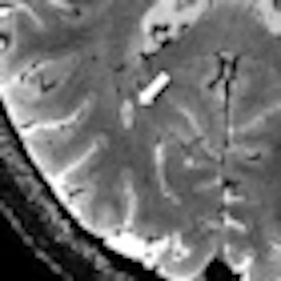
Using the 7-tesla MRI technique, 20 (80%) of the 25 patients could be diagnosed. Thirteen patients were diagnosed with MS; all of these patients had central veins visible in more than 40% of brain lesions. The seven patients with an eventual non-MS diagnosis had central veins visible in less than 40% of brain lesions.
 |
| Using 7-tesla and 3-tesla T2*-weighted MRI, multiple sclerosis lesions characteristically had appearances reminiscent of a donut or coffee bean. Using guidelines based on counting a few of these characteristic lesions, the diagnosis could be correctly identified. Images courtesy of Dr. Niraj Mistry. |
 |
The authors noted that lesions were found in all white-matter regions in the brain. They did not, however, look for gray-matter lesions in this study. Due to technical limitations, they could not scan the cerebellum, but Mistry said he and his colleagues would expect the results to be the same.
"These findings could lead to the first-ever single diagnostic test for the inflammatory demyelination that leads to MS," Mistry added. "It could also avoid further tests, such as spinal taps, and the diagnostic delay experienced by many patients suspected of having MS."
Unfortunately, 7-tesla MRI scanners are exceedingly rare outside the research setting. However, 3-tesla magnets are becoming increasingly common, and Mistry and colleagues are continuing their research by optimizing the T2*-weighted technique to work on these scanners.
"If our findings can be corroborated in a larger prospective 3-tesla MRI study, the technique could be rapidly transferred to healthcare settings," Mistry added.
The U.K. Multiple Sclerosis Society funded the study.