Women with newly diagnosed breast cancer should undergo a preoperative MRI scan to detect additional unsuspected malignancies, even if they do not have dense breasts, according to a study presented on Friday at the American Roentgen Ray Society (ARRS) annual meeting in Vancouver.
Researchers from Yale University in New Haven, CT, found that 3-tesla MRI detected a similar percentage of additional cancers and high-risk lesions in women with nondense breast tissue (26%) as in women with dense breast tissue (25%).
"We are seeing very comparable findings, so perhaps the mammographic breast density should not at all influence the decision to order an MRI," study co-author Dr. Reni Butler, assistant professor of diagnostic radiology, told AuntMinnie.com. Study results were presented by lead study author and breast imaging fellow Dr. Reena Vashi.
The authors initiated the study, in part, because there are currently no guidelines that define the role of breast density in determining if a preoperative MRI should be performed.
"We had made the anecdotal observation that our surgeons tend to refer patients with dense breasts more often, thinking that breast tissue can hide additional malignancies that we might be able to see better with MR," Butler said. "But they did not do as much [MRI] for patients who did not have dense breasts, thinking that the mammogram -- which we know is most sensitive if there is no dense breast tissue -- would be sufficient. So, we wondered if MRI would be helpful in those [nondense] patients as well."
Study population
The researchers evaluated 147 consecutive patients with newly diagnosed breast cancer over five months at their institution. Three-tesla MR images were obtained with a dedicated breast coil to help evaluate the extent of disease.
The group recorded each patient's age, breast density, additional suspicious lesions detected with breast MRI, and additional diagnosed malignancies and high-risk lesions. Patients were excluded if no recent mammogram was available to establish breast density, or if individuals were treated with mastectomy or neoadjuvant chemotherapy.
After exclusion criteria were considered, there were 127 patients in the study: 52 women who did not have dense breast tissue (41%) and 75 women with dense breasts (59%).
In their analysis, the researchers found a comparable percentage of additional malignancies in the two patient populations, with 26% of the patients without dense breasts having additional unsuspected malignancies, compared with 25% of the patients with dense breasts.
In addition, among women with dense breasts, 55% had additional suspicious lesions on 3-tesla MRI. Additional suspicious lesions were seen in 53% of the women with nondense breasts.
 |
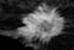
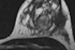
| Above, normal screening mammogram of contralateral right breast in patient with newly diagnosed left breast cancer and "not dense" breast tissue. Below, MRI reveals suspicious mammographically occult mass in right breast; MRI-guided biopsy yields diagnosis of contralateral infiltrating ductal carcinoma. All images courtesy of Dr. Reni Butler. |
 |
The researchers also identified suspicious mammographically occult lesions in 69% of the women in the nondense-breast category, while mammographically occult lesions were discovered in 71% of the women with dense breasts. High-risk lesions were detected in an additional 10% of nondense breast patients, and in an additional 11% of dense-breast patients.
Based on the results, Butler and colleagues concluded there was no statistically significant difference in the likelihood of detecting additional malignancies and high-risk lesions in newly diagnosed breast cancer patients on the basis of their mammographic breast density.
Butler said she and her colleagues were surprised by the results. "Our own anecdotal impression was that perhaps MRI might be useful in a few of those patients with dense breasts, but it was not as necessary," she added. "In this case, we are seeing very comparable findings, so perhaps the mammographic breast density should not at all influence the decision to order an MR."
When asked about the parameters for recommending an MRI scan after mammography to avoid unnecessary imaging, Butler said the results would indicate that MRI is helpful in almost every patient with newly diagnosed breast cancer.
"Sometimes women who are much older or who have other underlying conditions may not want to go through extensive surgery," she said. "Those patients may not benefit very much from finding these additional tiny cancers. There may be other medical issues that are more important."
Benefits of 3-tesla MRI
The stronger magnet strength of 3-tesla MRI also helps detect the additional malignancies. In a separate study, Butler and colleagues found that the upgrade to 3-tesla MRI naturally produced higher-resolution images.
"On the other hand, even with the 1.5-tesla magnet, [the results] are generalizable because comparing dense and nondense-breast patients would be useful in both patient populations," she said.
Butler and her colleagues plan to continue their research along this path. One of the limitations of the study is the small sample of only 127 patients, she noted.
"We would love to see a larger patient population to confirm these results," Butler added. "Also, we have very few patients in the extreme categories of predominantly fatty tissue or extremely dense tissue. We would like to see a broader spectrum and evaluate whether these results are consistent" in that patient population.