Italian researchers have found that one dose of gadobutrol appears to be just as effective as a double dose of gadopentetate dimeglumine for detecting late enhancement in cardiac MRI cases, potentially reducing possible adverse effects from contrast use.
The findings suggest that using less contrast could reduce the risk of patients developing nephrogenic systemic fibrosis (NSF), without negatively affecting image quality (American Journal of Roentgenology, April 2012, Vol. 198:4, pp. 809-816).
The study was led by Dr. Francesco De Cobelli, from the department of radiology and the Center for Experimental Imaging at San Raffaele Scientific Institute in Milan.
Contrast face-off
The researchers evaluated a 0.1 mmol/kg dose of gadobutrol (Gadovist, Bayer HealthCare Pharmaceuticals) and a 0.2 mmol/kg dose of gadopentetate dimeglumine (Magnevist, Bayer) in cardiac MRI patients between August 2008 and March 2010.
The 20 subjects included 14 men and six women with a mean age of 53 years, ranging from 20 to 81 years. All patients had previously undergone cardiac MRI with gadopentetate dimeglumine, with their images showing late enhancement.
The researchers excluded subjects with a history of allergic reaction to MRI contrast media, as well as those who were pregnant, unable to lie down for at least 45 to 60 minutes, or had contraindications to MRI, such as a pacemaker or claustrophobia.
Nine of the 20 patients had myocarditis, five had chronic coronary artery disease, and six had cardiomyopathy.
The mean time between the two MRI exams was 5.35 days. Late enhancement scans were acquired on a 1.5-tesla whole-body scanner (Achieva Nova, Philips Healthcare) on both short- and long-axis planes, with images acquired at 10-, 15-, and 20-minute intervals after gadobutrol injection. Total acquisition time averaged 40 minutes.
The MR images were evaluated on a two-point scale based on the presence or absence of late enhancement. Readers also measured signal-to-noise ratio and contrast-to-noise ratio based on signal intensities obtained from regions of interest in the normal myocardium, late enhancement areas, and left ventricular cavity.
Comparable results
The analysis found that late enhancement did not significantly differ between cardiac MRI with gadobutrol and gadopentetate dimeglumine, both in terms of total volume of myocardium (37.8 ± 56.1 cm3 and 35.1 ± 46.7 cm3, respectively) and percentage of involvement of the myocardial wall (22.5 ± 19.1% and 22.0 ± 17.2%, respectively).
Among the subgroups of patients with acute myocarditis, coronary artery disease, and cardiomyopathy, the researchers also found no significant difference in the performance of the two contrast agents.
Late enhancement detection for subgroups
|
||||||||||||||||||||||||||||||
The number of left ventricular segments visualized with the agents also showed no significant difference, with 138 segments for gadopentetate dimeglumine and 134 for gadobutrol.
There was little difference in other areas investigated by the researchers, as well. Signal-to-noise ratio did not differ significantly with gadopentetate dimeglumine (123.8 ± 82.9) versus gadobutrol (117.2 ± 88.6). Contrast-to-noise ratio was also similar between gadopentetate dimeglumine (96.2 ± 68.9) and gadobutrol (88.4 ± 72.9). In addition, contrast-to-noise ratio values between late enhancement and cavity signal did not differ significantly for the two agents.
 |
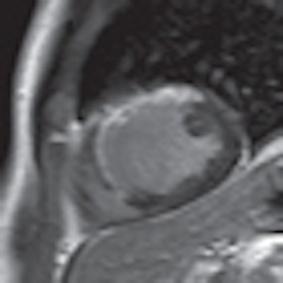
| MR images are of a 47-year-old man. Gadopentetate-enhanced image (left) shows large anterior transmural myocardial late enhancement, while corresponding gadobutrol-enhanced image (right) shows the same area, extension, and transmurality of infarct. Images courtesy of AJR. |
The significance of the study, according to the authors, is that late enhancement from single-dose gadobutrol for the first time was "not inferior" to results from double-dose gadopentetate dimeglumine. Their conclusion was supported by similar results in the pattern of contrast distribution, signal-to-noise ratio, and contrast-to-noise ratio.
"We believe that, combining the high efficacy in the cardiac perfusion in previously reported studies and our results of good performance on late enhancement depiction, gadobutrol may be considered an excellent choice for cardiac MRI," De Cobelli and colleagues concluded.
Gadopentetate dimeglumine has been the most-used gadolinium-based extracellular contrast agent for detecting late enhancement; however, use of this agent is now being questioned because of reports of NSF in patients with severe renal failure.
Limitations of the study included its size, with only a small group of patients with coronary artery disease, myocarditis, and cardiomyopathies evaluated.
Another potential limitation was allowing up to two weeks between the MRI scans with the two agents. The delay could potentially produce a time bias in the imaging of late enhancement, the authors wrote.
| The study was supported by a grant from Bayer Schering Pharma. |