British researchers used MRI to discover that the type of plaque in the carotid wall, rather than the extent of luminal stenosis, may be a better predictor of who will experience a minor stroke or transient ischemic attack (TIA), according to an April 17 study in the Journal of the American College of Cardiology.
By accurately assessing patients according to their lesion types, rather than the extent of luminal stenosis, the study from John Radcliffe Hospital in Oxford, U.K., suggests that correlating the MR images with brain injuries will allow for more effective treatment and therapy for patients earlier in the recuperative process. The lead study author is Dr. Alistair Lindsay from the hospital's Acute Stroke Service (JACC, Vol. 5:4, pp. 388-396.)
The researchers examined hospital records to look for patients who presented with a TIA or stroke at their facility. All study participants were diagnosed with a cerebrovascular event. To create a control group for comparison, asymptomatic patients were chosen for their degree of carotid stenosis, as well as age and sex, and compared to the symptomatic group.
Carotid artery imaging
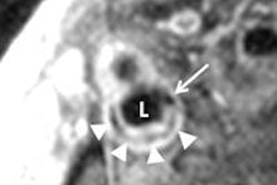
The patients' carotid walls were imaged on a 3-tesla MRI scanner with a phased-array, four-channel carotid coil. The researchers used a time-of-flight (TOF) MR angiography sequence to obtain bright-blood imaging of each carotid artery, selecting the artery contralateral to the side of clinical symptoms and bifurcating the artery to become a center landmark.
The researchers then followed with a brain MRI scan using a 12-channel head receiver coil. All patients underwent diffusion-weighted MR imaging (DWI) and fluid-attenuated inversion recovery (FLAIR) imaging of the brain.
Readers were blinded to the identification and clinical data for each patient. In the six cases in which a final interpretation differed, the classification was determined by consensus after further image review. MR images of the patients' plaques were graded on a scale of I to VI, according to American Heart Association standards.
Lindsay and colleagues found a total of 82 patients for the study. MR images were available from 41 symptomatic patients, as the 42nd symptomatic patient was unable to complete an entire MRI scan. Complete MR imaging data was obtained from all 40 patients in the asymptomatic group.
All symptomatic patients were diagnosed as having a stroke or TIA, with two physicians determining stroke in 24 cases and TIA for 17 individuals. The median time from symptom onset to MRI in the symptomatic group was 2.1 days, ranging from 0.2 to seven days.
Lesion evaluation
A review of the lesion types between the affected arteries and control arteries showed features consistent with hemorrhage, rupture, or thrombus. Those characteristics were seen in 22 of 41 (54%) symptomatic patients, but only eight of 40 (20%; p < 0.01) cases among the asymptomatic patients.
Significantly, Lindsay and colleagues noted, 12 of the 22 (55%) type VI plaques in the symptomatic group were in vessels with less than 70% stenosis of the lumen, as assessed by ultrasound.
A further analysis of the type VI plaque found surface rupture at all degrees of stenosis, including four cases causing less than 70% stenosis and two cases causing less than 50% stenosis of the carotid artery, as assessed by ultrasound. Surface rupture also was seen more often in symptomatic carotid arteries (24%), compared with asymptomatic arteries (5%).
Researchers also conducted follow-up MR imaging for the symptomatic group, with 30 of 41 (73%) subjects participating. Mean time to the follow-up MRI scan was 90 days, ranging from 30 to 250 days. The mean age of individuals having the follow-up MRI was 73.8 years, ranging from 48 to 90.6 years of age.
Plaque rupture
Of those 30 patients, evidence of acute plaque rupture was evident in 14 cases (46%) in the troubled artery on the initial MRI scan. Of the remaining eight type VI plaques scanned, six patients with type VI plaque characteristics were seen on the follow-up MRI. In two cases, there was evidence of the artery healing.
Results from diffusion-weighted MR images also revealed evidence of cerebral injury among 32 of 41 patients (78%) in the symptomatic group. The median number of lesions per patient was seven, with a median total lesion volume of 10.62 ml, ranging from 0 to 522 ml.
Researchers also found no lesions with diffusion-weighted MRI in the control group, and no significant associations in the control group between plaque characteristics and FLAIR signal.
In conclusion, Lindsay and colleagues wrote that the use of high-resolution 3-tesla MRI "to characterize carotid plaques of patients within seven days of minor stroke or TIA was feasible and showed a higher proportion of complex AHA type VI plaques compared with asymptomatic control patients."
In addition, more than 50% of complex plaques were found in arteries with less than 70% luminal stenosis, with plaque rupture associated with increases in both diffusion-weighted MRI and FLAIR lesions in the brain.
They added that the findings may provide a way to acutely assess patients according to their lesion types and not solely on the extent of luminal stenosis and to correlate those images with brain injury and determine more appropriate treatment and therapy.