Neuroimaging is an important tool to identify and evaluate traumatic head injury in victims of child abuse. Now, specific findings on CT and MRI can predict which patients have the highest risk for seizures, according to a study published online in Neurocritical Care.
Children with traumatic head injuries who experience clinical seizures tend to have poorer outcomes than children who are seizure-free. Pediatric specialists in Chicago believe that if patients at high risk for seizures can be identified upon hospital admission, more appropriate treatment protocols may be identified. They undertook a study to determine if clinically significant predictors of seizure could be associated with specific CT and MRI findings (Neurocritical Care, November 2, 2010).
A multidisciplinary team representing pediatric neurologists, radiologists, and critical care specialists at Northwestern University Feinberg School of Medicine in Chicago reviewed the medical records of all children diagnosed with abusive head trauma who were admitted to Children's Memorial Hospital in Chicago between January 2002 and March 2007.
A total of 54 children were identified. The majority were boys (61%) and infants (average age, 7.7 months). A total of 18 patients had one or more clinical seizures while hospitalized. This included nine patients who were among 21 individuals reported to have had seizures prior to hospital admission. For these nine patients, the researchers were unable to identify any significant association between prehospitalization seizures and seizures that occurred during the first week of hospitalization.
Fifty-two of the patients had head CT scans performed within the first 24 hours of admission to Children's Memorial Hospital, and 31 patients had a second CT scan during their first week. Additionally, 76% also had a brain MRI exam in this time period.
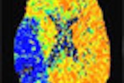
Subdural hemorrhages were identified in 90% of the CT exam findings, according to lead author Joshua Goldstein, MD, child neurology residency program director and assistant professor of pediatrics. Ninety-five percent of the MRI scans also identified subdural hemorrhages, and 31.7% showed an abnormal pattern of restricted diffusion.
The researchers were able to correlate midline shift, cerebral edema, and loss of gray-white differentiation on the initial CT scan images of patients who had subsequent seizures. MRI exam findings that were predictive of future seizures included midline shift, cerebral edema, infarction, and restricted diffusion. Six of the nine patients with midline shift present on the initial CT had clinical seizures, Goldstein reported.
Eleven of the 18 patients who had clinical seizures ultimately died from their injuries or required substantial inpatient rehabilitation.
By Cynthia E. Keen
AuntMinnie.com staff writer
December 29, 2010
Related Reading
CT fails to explain racial variation in child abuse mortality, April 9, 2010
Several clinical markers help detect abusive head trauma in young children, October 28, 2008
Copyright © 2010 AuntMinnie.com