People with a known high risk for Alzheimer's disease develop abnormal brain function even before the appearance of amyloid plaques that are characteristic of the disease, according to a new study published online December 15 in the Journal of Neuroscience.
Using functional MRI, researchers at Washington University School of Medicine in St. Louis determined that these patients have a particular form of the apolipoprotein E gene (APOE4). The findings suggest that the gene variant affects function long before the brain begins accumulating the amyloid that will eventually lead to dementia.
The lead author of the study was Yvette Sheline, MD, a professor of psychiatry, radiology, and neurology and director of Washington University's Center for Depression, Stress, and Neuroimaging.
The study involved functional MRI scans performed on 100 people with an average age of 62 years. Slightly less than half of the subjects carried the APOE4 variant, which is a genetic risk factor for Alzheimer's disease. Earlier PET scans of the subjects demonstrated that they did not have deposits in the brain of amyloid, the protein that contributes to plaque development in Alzheimer's patients and interferes with cognitive function.
Participants in the study also underwent spinal puncture tests that showed normal amyloid levels in their cerebrospinal fluid.
Sheline and colleagues found normal brain amyloid and cerebrospinal fluid levels in the subjects, but patients with the APOE4 variant had significant differences in the way various brain regions connected with one another.
In the study, subjects relaxed in an MRI scanner and reflected or daydreamed, while the machine measured oxygen levels and blood flow in the brain. Patients were given no tasks to perform but could not fall asleep, in order for the researchers to examine how the subjects' brains function at rest.
Although the researchers found no evidence of dementia or amyloid deposits, subjects with the APOE4 variant exhibited irregular functioning in the default mode network.
 |
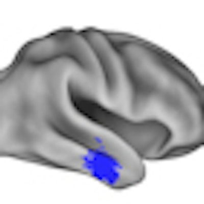
| Researchers identified functional differences in the brains of APOE4-positive and APOE4-negative subjects. Red areas indicate increased connectivity among regions at rest, while blue regions show decreased connectivity. Image courtesy of the Washington University School of Medicine. |
Sheline and colleagues speculated that a significant number of subjects eventually will be positive for amyloid. If some subjects do accumulate amyloid, the researchers will be able to look back at their data and identify particular patterns of brain function that may eventually be used to predict the onset of Alzheimer's disease.
By identifying patients with the highest risk of Alzheimer's, early treatment may begin to slow the progression of the disease and keep it from advancing to the stage when amyloid begins to accumulate and lead to dementia.
The research was supported by grants from the National Institute of Mental Health and the National Institute on Aging of the U.S. National Institutes of Health.
By Wayne Forrest
AuntMinnie.com staff writer
December 17, 2010
Related Reading
C-11 PiB-PET shows first progression of Alzheimer's disease, December 16, 2010
Walking reduces Alzheimer's progression, 1.5-tesla MRI shows, November 29, 2010
MRI shows physical activity's effect in preventing Alzheimer's, October 14, 2010
MRI helps identify mild cognitive impairment, October 5, 2010
MRI beats FDG-PET for early Alzheimer's detection, August 27, 2010
Copyright © 2010 AuntMinnie.com