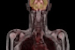
Fused PET and MR images are helping researchers better understand the long-term functional and structural changes that take place after traumatic brain injury, according to an animal study published in the November issue of the Journal of Nuclear Medicine.
By inducing fluid percussion injury in the brains of rats, researchers have found early widespread hypometabolic changes in certain regions and delayed progressive atrophic changes that continued to evolve for several months.
In particular, changes in the hippocampus, which is critical to memory and emotion, may have adverse implications for long-term neurological and psychiatric morbidity, which can follow traumatic brain injury, even when abnormalities are not obvious on structural MRI.
The lead author of the study was Ying R. Liu, from the department of medicine at the Royal Melbourne Hospital in Victoria, Australia (JNM, November 2010, Vol. 51:11, pp. 1788-1795).
Periodic imaging
The researchers noted that FDG-PET can map regional metabolism patterns in the brain in both humans and animals. Cross-sectional FDG-PET scans in humans after severe traumatic brain injury suggest a disruption in metabolic brain function.
FDG-PET scans were obtained from 16 rats that underwent fluid percussion injury, and from 11 rats that were part a healthy control group. Rats were injected with 37 to 74 MBq (1-2 mCi) of FDG 30 minutes before scanning on an animal PET scanner (Mosaic, Philips Healthcare, Andover, MA).
PET images were taken one week, one month, three months, and six months after injury, and they were acquired one to three days before MRI scans were taken of the same 11 regions-of-interest in the 27 rat brains.
T2-weighted small-animal MRI scans were obtained at baseline, one week, one month, three months, and six months after fluid percussion injury. MR images were acquired using a 4.7-tesla magnet (BioSpec, Bruker BioSpin, Billerica, MA) with a shield-gradient adjusted for rat imaging.
The rats also underwent several tests to assess anxietylike behavior and a test of learning and memory. Researchers used an open-field test and maze at one, three, and six months to gauge the rats' reactions and behavior after the induced injuries.
Image evaluation
The review of images showed differences between the healthy group and the injured rats in three regions-of-interest on the PET scans -- the left cortex, left hippocampus, and left amygdala.
In addition, the ipsilateral cortex showed significantly lower FDG uptake (17%) in injured rats three months after injury. FDG uptake also was reduced in the ipsilateral hippocampus at all time points, with a statistically significant difference at one week and three months.
In the ipsilateral amygdala, researchers found a significant reduction in FDG uptake in injured rats at one month and six months after the induced injury, compared to the healthy group.
 |
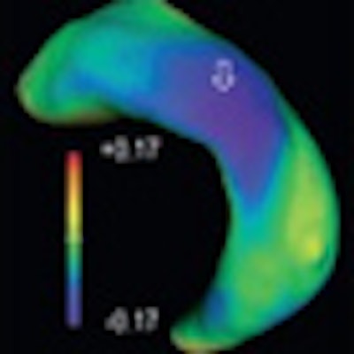
| Large-deformation high-dimensional mapping shows differences in left hippocampi between fluid percussion injury and healthy rats from baseline (far left) to six months (far right), with regional deformation distances for left hippocampi (ipsilateral to injury) in lateral and slightly caudal view. Open arrows show region of inward deformation (second from left) primarily representing cornu ammonis, which becomes visible at one week, progresses at one month (middle) and three months (second from right), and becomes less prominent at six months. Closed arrow at three months and six months shows regions of accentuated volume loss. Images courtesy of JNM. |
Behavioral differences
The researchers also found behavioral differences between the injured and healthy rats. The rats who had received the fluid percussion injury were more anxious and spent less time in an open-field test and maze.
"There was no confounding effect of locomotor activity in the fluid percussion injury group, because there was no significant difference in the distance traveled in the [maze]," Liu and colleagues wrote. "Similarly, fluid percussion injury rats spent less time and made fewer entries into the inner area of the open field, indicative of increased anxiety. Again, locomotor activity was not different between [fluid percussive injury] and sham rats in the open field."
Based on the rats' behavior and PET/MRI results, the researchers concluded that their study has "implications for the understanding of the pathophysiology and evolution of long-term neurologic morbidity following traumatic brain injury, and indicate an extended window for targeted neuroprotective interventions."
By Wayne Forrest
AuntMinnie.com staff writer
November 8, 2010
Related Reading
MRI detects white-matter brain damage in MS patients, November 2, 2010
MRI finds missed injuries on negative CT of blunt trauma, May 12, 2010
DWI-MRI predicts pediatric brain trauma outcomes, November 13, 2008
Diffusion-tensor MRI spots subtle changes after mild traumatic brain imaging, March 31, 2008
Copyright © 2010 AuntMinnie.com