Exercise is great for building muscles and bones, but what about the brain? With the help of high-resolution MRI, researchers at the University of California, Los Angeles (UCLA) have found that people who practice regular meditation have increased brain volume in certain regions.
The study, published the journal NeuroImage (April 2009, Vol. 45:3, pp. 672-678), found that certain brain regions in people who practiced meditation over a long-term period of time were larger than in a similar control group.
Lead author Dr. Eileen Luders, a postdoctoral research fellow at the UCLA Laboratory of Neuro Imaging, and colleagues said their work confirmed the beneficial aspects of meditation. In addition to having better focus and control over their emotions, many people who meditate regularly have reduced levels of stress and bolstered immune systems. However, less is known about the link between meditation and brain structure.
In the study, Luders and her colleagues examined 44 people: 22 control subjects and 22 individuals who had practiced various forms of meditation, including Zazen, Samatha and Vipassana, among others. The amount of time they practiced ranged from five to 46 years, with an average of 24 years.
More than half of the meditators said that deep concentration was an essential part of their practice, and most meditated between 10 and 90 minutes every day.
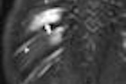
The researchers used high-resolution 3D MRI and two different approaches to measure differences in brain structure. One approach automatically divides the brain into several regions of interest, allowing researchers to compare the size of certain brain structures. The other approach segments the brain into different tissue types, allowing researchers to compare the amount of gray matter within specific regions of the brain.
The researchers found significantly larger cerebral measurements in meditators compared with controls, including larger volumes of the right hippocampus and increased gray matter in the right orbitofrontal cortex, the right thalamus, and the left inferior temporal lobe. There were no regions where controls had significantly larger volumes or more gray matter than meditators.
Because these areas of the brain are closely linked to emotion, Luders said these regions might be the neuronal underpinnings that give meditators the ability to regulate their emotions and allow for well-adjusted responses to life's occurrences.
Related Reading
Illuminating the mind: Imaging explores the mysteries of meditation, September 11, 2003
Meditation shown to light up brains of Buddhists, May 23, 2003
Strike a pose: PET sheds light on benefits of yoga, December 31, 2002
Copyright © 2009 AuntMinnie.com