By combining two MR imaging techniques to view gray and white brain matter in active professional fighters, researchers can identify and track the progression of cognitive impairment, according to a study published online July 25 in Radiology.
T1-weighted MRI and diffusion-tensor imaging (DTI) derived from diffusion-weighted MRI revealed a set of seven imaging predictors for cognitive function among more than 250 fighters. Lower gray-matter volume and thickness, along with lower white-matter tract integrity that declined over time, collectively predicted which fighters would be cognitively impaired, according to the team led by Virendra Mishra, PhD, from the Cleveland Clinic Lou Ruvo Center for Brain Health in Las Vegas.
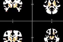
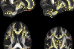
DTI is specific to the white-matter part of the brain and T1-weighted images are sensitive to the gray matter, Mishra said. By combining these approaches, the researchers hoped to find imaging biomarkers on MRI that could be used to predict whether or not fighters will become impaired.
Mishra and colleagues collected data from the Professional Fighters Brain Health Study, which is one of the largest longitudinal studies of brain health in contact sports. The information included 273 male fighters who had undergone baseline T1-weighted MRI and DTI scans and 56 fighters who returned for a follow-up scan. The subjects were given neuropsychological tests to assess their processing and psychomotor speeds and to differentiate between fighters with cognitive impairment and those who were not impaired.
The researchers found an association between regions of gray matter and white matter and the fighters' cognitive function. For example, T1-weighted volumetric measurements of the left thalamus, which acts as a relay center to connect a multitude of brain regions, helped distinguish cognitively impaired and nonimpaired fighters. In addition, fractional anisotropy values, which reflect white-matter integrity, along two different white-matter tracts were identified as possible predictors of cognitive impairment. In all, the researchers tagged seven brain regions composed of gray and white matter that could become imaging biomarkers for cognitive impairment.
This MRI approach may also have other applications, such as evaluating the efficacy in clinical trials of novel therapeutics designed to reduce the risk of cognitive impairment. It could also be used to study other contact sports in which head injuries and mild traumatic brain injury (TBI) are common. In addition, the group is interested in the order of the effects of repeated trauma: whether neurons in the gray matter or the fiber tracts in the white matter are affected first.