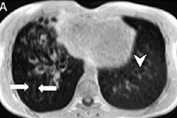
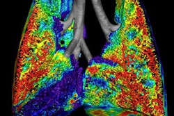
Researchers from the University of Missouri (MU) are combining MRI and a form of helium used as a contrast agent to visualize lung function in people with cystic fibrosis.
The contrast-enhanced MRI scans would be used to image patients with a gene mutation known as G551D-CFTR, which causes a salt imbalance due to the defective cystic fibrosis transmembrane conductance regulator (CFTR) protein. Patients with the condition take the drug ivacaftor, but the efficacy of the treatment is not well-understood, said lead author Dr. Talissa Altes, chair of the radiology department at MU School of Medicine, in a statement.
The two-part study investigated the use of MRI with helium-3 as a contrast agent to evaluate lung function. In the first part, eight patients were given either ivacaftor or a placebo for four weeks to measure short-term effectiveness. In the second part, nine patients received ivacaftor for 48 weeks to determine the drug's long-term efficacy. After each phase, patients had a spirometry test to measure their ability to breathe and underwent MRI with the hyperpolarized helium (Journal of Cystic Fibrosis, March 2017, Vol. 16:2, pp. 267-274).
"We found that after using ivacaftor, patients experienced a dramatic increase in lung improvement in both the short and long term," Altes said. With more study, the researchers hope to use helium-3 MRI in younger children or babies with impaired lung function or other respiratory diseases.