Researchers have developed a new cardiac PET/MRI protocol with sodium fluoride (NaF) that significantly reduces radiation dose and artifacts, enhancing the view of disease activity in coronary arteries.
Using the protocol greatly improves image quality, and patients benefit with less radiation exposure from PET/MRI compared to cardiac PET/CT scans, according to the study presented earlier this month at the American College of Cardiology (ACC) annual meeting in Chicago.
"This new sequence does a much better job of segmenting out the different tissues in the body so we can get accurate attenuation correction maps and account for the effects of cardiac motion, which is the other source of artifacts," said study co-author Dr. Marc Dweck, British Heart Foundation senior lecturer at Mount Sinai's Icahn School of Medicine. "With low-radiation scans of less than 5 mSv, we can get very interesting information about disease activity within the heart."
Cardiac PET/MRI
Mount Sinai has been performing and researching PET imaging of the heart for many years. One of the hospital's initiatives is to assess the uptake of various tracers, predominantly FDG and sodium fluoride, in atherosclerosis and aortic stenosis. Likewise, PET/CT has been a staple of the facility's cardiology department and is used to investigate myriad cardiac conditions.
Of course, in recent years, radiation exposure to patients from PET/CT has become a primary concern among clinicians, cardiologists, and other practitioners.
PET/MRI, meanwhile, has been in place at Mount Sinai for about five years now, with much of the hybrid modality's work targeting large vessel, aorta, and myocardial imaging, Dweck said.
"If we are going to consider using these [modalities] for clinical applications, then it would be nice to have the radiation doses as low as possible," he told AuntMinnie.com. "So, PET/MRI is something we have been keen to explore for a long time."
Since he arrived at Mount Sinai about 10 months ago, the facility has used PET/MRI on more than 70 patients for a wide range of maladies, including myocardial disease, cardiac sarcoidosis, and the presence of amyloid plaque. About half of the scans focused on the coronary arteries, Dweck estimated.
PET/MRI "is a modality that is in its infancy, particularly in respect to cardiovascular applications, but we were keen initially to look at a wide range of conditions and see where it may be useful," he added.
NaF's utility
For cardiac studies, the tracer sodium fluoride is used to gather information on calcifications, as opposed to FDG, which provides images of inflammation. The primary advantage of NaF is that there's no uptake of the tracer by surrounding tissues.
"You can get excellent signal-to-noise in the coronary arteries, in the aortic valve, and with aortic stenosis [with NaF]," Dweck explained. "FDG is hampered by relatively low signal and uptake in the adjacent myocardium."
In the current study, eight patients diagnosed with cardiovascular disease or related risk factors were imaged with simultaneous PET/MRI (Biograph mMR, Siemens Healthcare). PET and MRI data were acquired 40 to 90 minutes after injection of 18 mCi of sodium fluoride.
The MR attenuation correction maps were calculated in one of two ways. One method was the standard vendor-recommended approach, which employed a 3D gradient-echo volumetric interpolated breath-hold examination (VIBE) sequence and was later segmented into four tissue classes based on background air, lung, soft tissue, and fat, and assigned linear attenuation values.
The second method was the Mount Sinai technique, which is a seven-minute, free-breathing, golden-angle radial gradient-echo VIBE sequence. Data acquired with the protocol were later segmented into background air and soft tissue based on image intensity. Bone was included in the calculations based on segmentation of its high NaF signal.
The term "golden angle" refers to a mathematical principle that states that a path moving at a certain angle around a circle will never cross the same point in that circle twice.
"The great advantage is that is if you are acquiring MRI data according to that principle, you are always acquiring new data and it is helping to build the MR image," Dweck explained. "It reduces scanning time, increases efficiency, and does not repeat work."
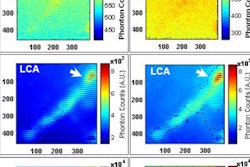
In analyzing the resulting images from the two MR attenuation correction methods, Dweck and colleagues found that the standard MR sequence produced a very bright and problematic artifact in the bronchus, the main conduit of air into the lungs, which is located very close to the heart.
By comparison, the Mount Sinai technique eliminated the visual distractions.
"The images were not dominated by the artifact; they were dominated by the actual [NaF] signal," Dweck said. "Without this advanced attenuation correction sequence, it is not feasible to look at coronary uptake [of NaF] and uptake in the heart muscle."
 Images show how Mount Sinai's MR attenuation correction technique (right column) eliminates artifacts (arrows) compared with standard attenuation correction (left column, arrows). Images courtesy of Dr. Marc Dweck.
What's next?
Dweck speculated that this novel MR attenuation correction technique with PET/MRI's lower radiation dose could encourage expanded use of the hybrid modality for testing new radiopharmaceuticals.
"With PET/MRI, you could look at two, three, four, or more time points with a radiation dose of, say, 20 mSv," he said. "With PET/CT, you are limited to only one or two time points. This is an exciting approach if you are conducting those kinds of studies."
Dweck also theorized about the use of both NaF and FDG with PET/MRI to assess inflammation and calcification at different time points of a scan.
"It could open up the ability to do some quite sophisticated imaging," he added.