Chinese researchers using diffusion-tensor MRI (DTI-MRI) have found abnormalities in the neural white-matter tracts of patients with insomnia, according to a study published online April 5 in Radiology.
While insomnia's adverse effects, which include daytime fatigue, mood disruption, and cognitive impairment, are well-known, the sleep disorder's causes are less clear.
So senior author Dr. Guihua Jiang and colleagues from Guangdong No. 2 Provincial People's Hospital in Guangzhou sought to analyze white-matter tracts with DTI and the relationship between abnormal white-matter integrity and the duration and features of insomnia. DTI is an MRI technique that enables radiologists to analyze the pattern of water movement along white-matter tracts to identify a loss of tract integrity.
Jiang and colleagues also used a technique known as tract-based spatial statistics, which is highly sensitive to the microstructure of the white-matter tract and provides multiple diffusion measures.
The study included 23 patients with primary insomnia and 30 healthy control subjects. All 53 participants completed questionnaires including the Pittsburgh Sleep Quality Index, the Insomnia Severity Index, the Self-Rating Anxiety Scale, and the Self-Rating Depression Scale to evaluate mental status and sleep patterns (Radiology, April 5, 2016).
The researchers found significantly reduced white-matter integrity in the insomniacs in several right-brain regions and the thalamus, which regulates consciousness, sleep, and alertness, compared with the healthy controls. In addition, abnormalities in the thalamus and body corpus callosum were associated with the duration of patients' insomnia and their self-rating depression scale score.
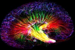
 DTI shows the distribution of the six whole white-matter tracts in the brain. ALIC = anterior limb of the internal capsule, ACR = anterior corona radiata, BCC = body of the corpus callosum, PLIC = posterior limb of the internal capsule, R = right side of the brain, SCR = superior corona radiata, SLF = superior longitudinal fasciculus. Image courtesy of RSNA.
Finding the role of the thalamus in insomnia is particularly critical, because this region of the brain is associated with the body's biological clock, according to lead author Shumei Li. The results also indicate that the underlying cause of white-matter integrity abnormalities in insomnia patients may be a loss of myelin, the protective coating around nerve fibers.
Jiang and colleagues recommended additional research with a larger patient sample to clarify and validate a link between altered white-matter integrity and insomnia.