Researchers using functional MRI (fMRI) have detected differences in the brains of children with autism spectrum disorder who are overly sensitive in terms of how they react to external stimuli, according to a study published on June 10 in JAMA Psychiatry.
The group from the University of California, Los Angeles (UCLA) used fMRI to image brain response to auditory and tactile stimuli in children and adolescents ages 9 to 17 with and without autism spectrum disorder. Some of the autistic subjects also had a condition called sensory overresponsivity (SOR), which affects more than 50% of children with autism.
Brain imaging research on SOR is still relatively new, and sensory symptoms were only recently added to the diagnostic criteria for autism. The condition can be debilitating for children with autism and their families, who often feel confined to their homes.
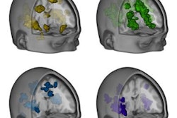
The researchers found that there were big differences in brain response between autistic kids with SOR and those with only autism -- the latter had brain responses more typical of kids without autism.
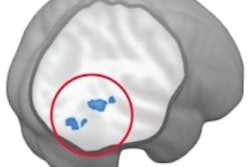
The responses were concentrated in brain areas that process sensory information -- the primary somatosensory and auditory cortices -- as well as in the amygdala, which is one of the brain's emotional centers. Exposing the individuals with SOR to both auditory and tactile stimuli simultaneously elicited even more severe responses.
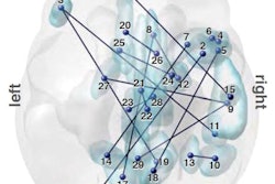
First author Shulamite Green, PhD, postulated that children with autism but not SOR might be compensating through strong brain connectivity between the prefrontal cortex and amygdala. The prefrontal cortex could be helping to regulate the strong response of the amygdala.
The findings indicate that SOR treatment could focus on creating coping skills to enable these children to handle stimulating environments, rather than changing sensory processing. In future work, the group hopes to better characterize the neurobiology of these patients' reactions.