A new MRI technique called restriction spectrum imaging MRI (RSI-MRI) is providing enhanced details on the extent and exact location of prostate tumors.
RSI-MRI was developed at the University of California, San Diego (UCSD) and the University of California, Los Angeles (UCLA), and the technique could lead to more precise and effective treatment for prostate cancer, according to a study published online January 6 in the journal Prostate Cancer and Prostatic Disease.
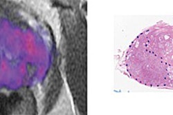
Led by senior author Anders Dale, PhD, professor of radiology at UCSD, the study details how RSI-MRI corrects for distortion caused by magnetic field artifacts from standard contrast-enhanced MRI. Dale and colleagues used RSI-MRI to identify the extension outside the prostate of a tumor in eight of nine patients compared with standard MRI, which found only two of the nine patients as having the extension.
The finding is significant, researchers explained, because the extension indicates that the tumor has grown beyond the boundary of the prostate into surrounding tissue, which, in turn, indicates a more severe form of the disease that requires more aggressive treatment.
Dale and colleagues added that additional clinical research is needed with a larger group of patients, but the results are encouraging, and RSI-MRI may be a valuable tool to identify patients with less severe disease and spare them unnecessary treatment.