Using an MRI technique called magnetic field correlation (MFC), researchers can measure brain iron levels in adolescents, which could help diagnose attention-deficit/hyperactivity disorder (ADHD) and lead to more effective treatment. Study results were published online June 17 in Radiology.
Magnetic field correlation was introduced in 2006 by study co-authors Joseph Helpern, PhD, and Jens Jensen, PhD, from the Medical University of South Carolina (MUSC). MFC is designed to measure iron levels through a decrease in MRI signal. As iron levels increase, there is a corresponding decrease in the MRI signal, which creates dark spots on MR images.
In a study first presented at RSNA 2013, the researchers measured brain iron levels in 22 children and adolescents with ADHD and 27 healthy controls. Subjects ranged in age from 8 to 18 years old. Twelve of the ADHD subjects had never used medication for their condition.
All subjects were imaged on a 3-tesla scanner (Magnetom Trio, Siemens Healthcare) to create the MFC images. No contrast agents were used, and blood iron levels in the body were measured using a blood draw (Radiology, June 17, 2014).
The results showed that the 12 patients who had never used ADHD medication had significantly lower striatal and thalamic MFC indexes of brain iron than the ADHD patients who had been on psychostimulant medication and the control group.
Conversely, the medicated ADHD patients had brain iron measures comparable to the controls, suggesting that brain iron may increase to normal levels with psychostimulant treatment.
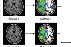
 MRI (far right) shows regions of interests (green) in the caudate nucleus (CN), putamen (PUT), globus pallidus (GP), and thalamus (THL) for controls, medication-naive ADHD patients, and ADHD patients with a history of psychostimulant treatment. The ADHD-nonmedicated subgroup displayed significantly reduced striatal (CN, PUT) and thalamic MFC compared to both controls and the ADHD-medicated subgroup. No significant differences were detected between the latter two groups. Image courtesy of Radiology.
The findings suggest that iron absorption in the brain may be abnormal in patients with ADHD, given that atypical brain iron levels are found even when blood iron levels in the body are normal, said lead author Vitria Adisetiyo, PhD, postdoctoral research fellow at MUSC.
Currently, ADHD diagnosis is based on subjective clinical interviews and questionnaires. If brain iron could serve as a noninvasive biomarker, this could help inform clinical diagnosis, particularly in borderline cases, Adisetiyo said.
MFC could have a future role in determining which patients would benefit from psychostimulants if the current results can be replicated in larger studies. According to the American Psychiatric Association, ADHD affects 3% to 7% of school-age children.