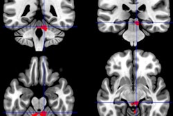
Using functional MRI (fMRI), researchers may have found two distinct forms of Gulf War illness based on signs of atrophy in different brain regions.
The findings may explain why veterans with Gulf War illness have reported different symptoms and complaints, according to a study published online June 14 in PLOS One.
Researchers at Georgetown University Medical Center used fMRI before and after exercise tests to study the effects of physical stress on the veterans and a control group.
In 18 veterans, they found that pain levels increased after completion of the exercise stress tests. In these subjects, fMRI showed the loss of brain matter in adjacent regions associated with pain regulation.
During cognitive tasks, this group showed an increased use of the basal ganglia, a compensation mechanism also seen in neurodegenerative disorders such as Alzheimer's disease. Following exercise, subjects in this group lost the ability to use their basal ganglia, suggesting an adverse response to a physiological stressor.
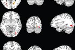
A separate group of 10 veterans showed a very different response, noted lead author Rakib Rayhan, a researcher in the lab of senior investigator Dr. James Baraniuk, professor of medicine at Georgetown.
In these 10 veterans, the researchers found substantial increases in heart rate, along with atrophy in the brain stem, which regulates heart rate. In addition, brain scans during a cognitive task performed prior to exercise showed increased use of the cerebellum, which, again, is seen in neurodegenerative disorders. Like the other group, this cohort lost the ability to use this compensatory area after exercise.