The combination of electroencephalography (EEG) and functional MRI (fMRI) leads to more precise localization of areas that cause epileptic seizures, giving neurosurgeons a better understanding of how to treat the condition, according to a study presented this week at the 2013 Canadian Neuroscience Meeting in Toronto.
Seizures can often be controlled using medication; however, in an estimated 40% of patients, drugs do not control seizures well. For some of these patients, surgery to remove the abnormal brain cells causing the seizures can be considered.
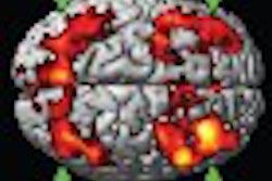
Before brain surgery can be performed, doctors and surgeons must determine the point of origin of the seizures, which can be viewed as brain activity spikes, and if cells can be removed without damaging other areas of the brain.
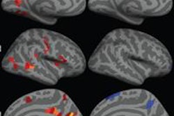
Dr. Jean Gotman and colleagues from McGill University's Montreal Neurological Institute combined EEG, which provides information on the timing of the spikes but cannot always pinpoint location, with fMRI, which can better identify the location.
EEG-fMRI can be technically challenging because fMRI's strong magnetic field can interfere with the recording of the electrical currents emitted by brain cells. However, the two modalities can be useful for patients in whom precise localization of the seizure origin is difficult.
"Combing EEG and fMRI is a unique method to define noninvasively in the whole brain the regions involved in epileptic discharges," the authors wrote. "It is a complex tool, but it is likely to play an increasing role among the methods currently used to localize the source of epileptic activity."