Using MRI, researchers at NYU Langone Medical Center believe they have taken an important first step in understanding the rotator cable's role in both intact and torn rotator cuffs, which, in turn, could improve characterization of a patient's injury or disease.
The study, published in the May issue of the American Journal of Roentgenology, concluded that by knowing the typical location and morphology of the rotator cable, physicians can better diagnose and treat conditions involving the cable, such as rotator cuff tears.
This improved characterization could also lead to additional studies correlating cable pathology with rotator cuff abnormality such as fatty atrophy, tear size, and tear retraction, wrote Dr. Soterios Gyftopoulos, from NYU Langone's department of radiology, and colleagues (AJR, Vol. 200:5, pp. 1101-1105).
Maintaining shoulder strength
The rotator cable becomes increasingly important as a person ages, helping to stabilize the rotator cuff tendons, the authors noted. When the cable is intact, the rotator cuff can maintain a person's shoulder strength and range of motion even when there is some tearing of atrophied fibers.
"If the cable became compromised, then the cuff would lose its main stabilizing structure, leading to progressive, worsening shoulder symptoms," the authors wrote.
Gyftopoulos and colleagues state "there is no consensus in terms of the MRI appearance of the rotator cable." Hence, the purpose of the current study was to define the characteristics of the rotator cable through MRI.
Over a two-week period, 41 male and 29 female patients (mean age, 45 years; range, 3 to 84 years) with intact rotator cuff tendons underwent MRI scans of their shoulders. Of the 70 scans, 34 were conducted on a 1.5-tesla MRI scanner and 36 were done on a 3-tesla system (both from Siemens Healthcare).
Two musculoskeletal radiologists independently reviewed each study and recorded the presence, location, and size of the patients' rotator cables. The researchers defined intact tendons as those with no evidence of partial-thickness or full-thickness tearing.
The rotator cable was visualized in 74% of the MRI studies on both coronal and sagittal oblique sequences by both readers. The mean width of the cable was 1.24 ± 0.31 cm and the mean thickness was 0.19 ± 0.05 cm.
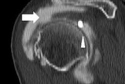
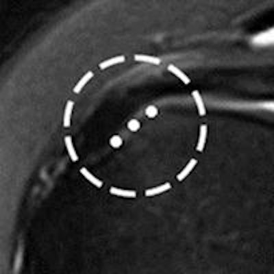
 The location of the cable was measured from medial enthesis to its lateral margin (circle) All images courtesy of AJR.
 Cable thickness was measured from its superior to inferior margins (circle).
 Width was measured from its medial to lateral margins (circle).
The mean cable thickness in this study was smaller than the 0.47 cm reported in previous research, the authors noted.
"This was most likely because the original measurements [in the previous study] included the adjacent joint capsule and rotator cuff tendon fibers in addition to the cable," Gyftopoulos and colleagues explained.
The rotator cable can be consistently seen on anatomic and histologic analysis, arthroscopy, and MRI of the intact rotator cuff, the group concluded.
"The realization that the rotator cable can be frequently seen on conventional MRI represents an important first step in understanding its possible biomechanical role in the intact and torn rotator cuff by use of imaging," they wrote.
The authors did acknowledge several limitations of the study, including its retrospective nature and the lack of surgical correlation.