In results that add substantial forensic power to identifying the cause of death, postmortem cardiac 3-tesla MRI has been found to identify sudden cardiac death in cases that are invisible at conventional autopsy, according to new research published online in the Journal of the American College of Cardiology.
In 76 cases, unenhanced MRI was able to visualize and discriminate 124 myocardial lesions, and there was excellent correlation among autopsy findings on chronic, subacute, and acute cases at MRI, the study authors said.
Thus, 3-tesla MRI can visualize chronic, subacute, and acute myocardial infarction in situ, and shows a possible source for sudden cardiac death in peracute infarction, wrote Dr. Christian Jackowski from the University of Bern in Switzerland, along with colleagues from the University of Lausanne in Switzerland and Linköping University in Sweden.
In particular, peracute infarction findings cannot be replicated in a physical autopsy; therefore, the study findings supports the use of forensic autopsy by permitting targeted histology, the group reported. Thus, MRI may serve as an alternative postmortem examination technique to traditional autopsy, Jackowski and colleagues wrote (JACC, April 3, 2013).
"In forensics, the cases can be solved better and more convincingly," Jackowski wrote in an email to AuntMinnie.com. "The second impact I see is for clinical pathology, which has suffered from declining autopsy rates for decades now. I don't expect the clinical autopsy numbers to increase again, so postmortem MRI can provide information about the cause of death, especially in patients that do not undergo a clinical autopsy anymore."
Most cardiac deaths remain unsolved
Considering that cardiac events account for most deaths, postmortem MRI's ability to demonstrate the cause of death in the heart has an enormous potential benefit, the group wrote, noting that "tissue alterations occurring during and after myocardial ischemia" are the most important -- yet the least understood -- issue with regard to conventional autopsy.
In previous small studies, acute, subacute, and chronic infarction have been differentiated based on signal behavior in T1- and T2-weighted images. Previous research has also suggested that unenhanced T2-weighted images without a hyperintense margin may represent acute ischemic lesions, which are aged between about 15 minutes and one hour, the group noted. Generally, though, cases of sudden cardiac death without myocardial findings of fresh thrombus have puzzled examiners and pathologists for decades.
The study sought to validate previous findings using postmortem MRI at 1.5 tesla, and also sought to use a larger study population. A secondary goal was to link the MRI finding of T2-weighted myocardial hypointensity to the possible myocardial appearance of sudden death to the patient's coronary status at autopsy. The group used 3-tesla MRI to examine 136 corpses whose case histories showed chronic or acute cardiac anamnesis, or death from circumstances suggesting a cardiac origin.
All cases were examined with MRI using acquired short-axis and horizontal long-axis imaging on a 3-tesla MRI system (Achieva, Philips Healthcare) using a 16-channel torso coil.
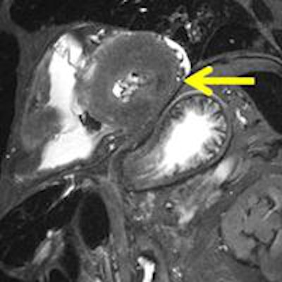
 Peracute myocardial infarction in postmortem MRI (death within one hour). A T2-weighted short-axis image presents with a local hypointensity within the lateral wall without hyperintense edematous margin. Autopsy of the specimen showed no visible alteration within the affected myocardium (not shown). Histology also failed to demonstrate ischemic alterations (not shown). Dissection of the coronary artery system revealed a fresh soft-plaque rupture with intimal hemorrhage within the circumflex coronary artery (not shown). Image courtesy of Dr. Christian Jackowski.
A total of 76 cases (62 men, 14 women; mean age at death 57.8 ± 16.7) presented with cardiac findings at postmortem MRI were investigated further.
Findings of cloudy hypointense myocardial areas in T2-weighted images without any hyperintense marginal edematous reaction were diagnosed as peracute ischemic lesions, and were not visible at autopsy, Jackowski and colleagues wrote.
Among the 76 cases, postmortem analysis identified 124 myocardial lesions (chronic = 25, subacute = 16, acute = 30, and peracute = 53), the team wrote. They found excellent correlation among myocardial findings at autopsy and chronic, subacute, and acute infarction cases. Peracute infarction areas detected at postmortem MRI were verified by histology in 62.3% of cases, and could be related to a matching coronary finding in 84.9%, they wrote.
In 15.1% of peracute lesions seen at MRI, the researchers found no matching coronary finding. But these patients presented severe myocardial hypertrophy or cocaine intoxication that "facilitated a finding of cardiac death without a verifiable coronary stenosis," they noted.
Postmortem 3-tesla MRI revealed chronic, subacute, and myocardial infarction, the authors concluded, and in cases of peracute infarction, MRI pinpoints the possible source of sudden cardiac death, demonstrating affected myocardial areas at autopsy, Jackowski and colleagues wrote.
More precision in defining cardiac death
The study is the first to amass a large number of cases presenting with hypointense T2-weighted lesions that are well-correlated to coronary events; among the 42 cases, there were 53 hypointense T2-weighted lesions that remained invisible at macroscopic dissection, the authors wrote.
"Knowing about the postmortem MR finding allowed for a targeted histological examination that showed early ischemic alterations in 62.3% of lesions," they wrote. "Based on the present study, it is ... expected that ischemic alterations would have not been detected in a routine histological examination without having the MR finding in advance."
At MRI, no histological alteration could be seen within the affected myocardium in 37.7% of the cases, the authors cautioned. On the other hand, a comparison of hypointensity in MRI to coronary status at autopsy showed findings that were able to explain an ischemic situation within the myocardium in 84.9% of the cases, demonstrating solid value for the postmortem imaging technique. In daily practice, this suggests that MRI could explain the majority of unsatisfactory autopsy cases thought to be sudden cardiac death but without verifiable myocardial changes.
Among the study limitations cited by the authors, the design did not permit the use of a control group, and the researchers were not blinded to the results, they stated, adding that the gold standard, autopsy, has several known limitations that hamper comparison with imaging.
Postmortem cardiac imaging comes with the disadvantage of no late enhancement. But it may be more than compensated for by the lack of cardiac motion or breathing artifacts.
Before this study, "we have seen hearts that presented with older or no ischemic lesions and may have three-vessel disease, but no fresh lesion that could explain why the heart stopped working exactly at that time," Jackowski told AuntMinnie.com. "We then speak about sudden death, meaning that an arrhythmic event might have caused the death. However, what causes the arrhythmia often remains unclear. With postmortem MRI, we do see myocardial areas that the arrhythmia possibly originates from, areas that remain hidden for the human eye at autopsy. In forensics, the cases can be solved better and more convincingly."