By using a new MRI technique to measure the wall thickness of coronary arteries, researchers from the U.S. National Institutes of Health (NIH) may be able to identify coronary heart disease early in its progression, according to a study published October 9 in Radiology.
Earlier detection of plaque and atherosclerosis can lead to earlier treatment for coronary artery disease and better patient outcomes. The problem is that coronary arteries are extremely difficult to image clearly because of their small size and constant motion, noted lead researcher Khaled Abd-Elmoniem, PhD, a staff scientist in the Biomedical and Metabolic Imaging branch of NIH's National Institute of Diabetes and Digestive and Kidney Diseases.
MRI could be used to image the coronary arterial wall, but there are technical challenges due to heart motion that can reduce the modality's success rate in coronary imaging. The failure rate of MR imaging of the coronary wall is 26% to 33%, compared with a failure rate of 10% to 20% for coronary MR angiography.
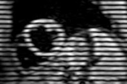
Two other challenges in 2D coronary wall artery imaging are that the section to be imaged is orthogonal to the longitudinal view of the artery at the location of interest, and that signal from blood be nulled. To that end, the authors tested a phase-sensitive dual inversion-recovery (PS-DIR) black blood imaging technique called time-resolved acquisition of PS-DIR (TRAPD).
The study evaluated 12 healthy individuals and 26 people with at least one coronary artery disease risk factor by using MRI to measure coronary artery wall thickness. The mean age of the 13 men and 13 women with known risk factors was 48 years, while the mean age of the three men and nine women who comprised the healthy control group was 26 years.
With TRAPD imaging, five continuous image frames are acquired to increase the ability to obtain a clear image with no blurring. Researchers found that the technique achieved a success rate of 95% in acquiring a good- to excellent-quality image, compared with 76% of the time with single-image dual inversion recovery.
Abd-Elmoniem and colleagues also found that the TRAPD-MRI resulted in a significant difference between wall thickness measurements of coronary artery disease (CAD) patients and healthy subjects and a smaller standard deviation, which is indicative of more precise measurements. Using TRAPD, subjects with CAD risk factors had vessel wall thickness measurements of 1.07 mm, compared with a thickness of 1.46 mm for those with no risk factors, a difference of 36%. The difference between the two groups was only 25% for the conventional MRI technique (1.24 mm versus 1.55 mm).
The results suggest that MRI may be used in the future to screen for individuals at risk for coronary artery disease, and may be useful for monitoring the effects of therapies, Abd-Elmoniem said. In addition, unlike blood tests that measure cholesterol and lipids in the blood, which can indicate atherosclerosis, coronary artery wall thickness is a direct measurement of early-stage coronary artery disease.
Abd-Elmoniem colleagues added that more research is needed to validate the TRAPD technique.