MRI has shown that changes in brain blood flow associated with vein abnormalities are not specific for multiple sclerosis (MS) and do not contribute to the affliction's severity, according to a study published online August 21 in Radiology.
Previous research has speculated that multiple stenoses restricting blood flow in the brain -- known as chronic cerebrospinal venous insufficiency (CCSVI) -- may contribute to MS; however, the findings from researchers at the University of Rome "Tor Vergata" contradict this theory.
The prospective study by Dr. Francesco Garaci, from the university's department of diagnostic imaging, and colleagues enrolled 65 subjects (41 women and 24 men; mean age, 42 years) from February to September 2011. Of this group, 39 subjects had MS and 26 were matched healthy controls who had no evidence of neurological disease (Radiology, August 21, 2012).
Inclusion criteria for the study included a clinical diagnosis of MS, with a relapsing-remitting or secondary progressive disease course. Subjects were excluded if they could not comply with the protocol, had contraindications to MRI, had a clinical history of cerebral thrombosis, or had undergone head and neck surgery. They were also excluded if they had experienced a relapse of MS in the 30 days prior to the start of the study.
Based on color Doppler ultrasound results, 25 of the MS patients were positive for CCSVI and 14 were negative. Among the healthy controls, 14 were positive for CCSVI and 12 were negative.
 |
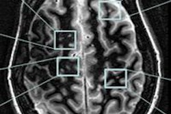
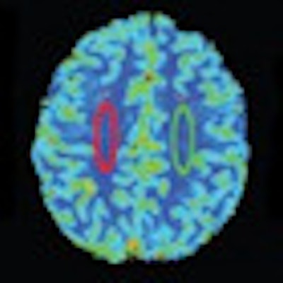
| Regions of interest (ROI) were placed in the white matter of a 38-year-old healthy subject at the level of the semioval centers to avoid arterial and venous structures. ROIs were in the same position and same measurements in all patients with MS and all control subjects. T2* = T2*-weighted MRI; CBV = cerebral blood volume; CBF = cerebral blood flow. Images courtesy of Radiology. |
The researchers used contrast-enhanced MRI to assess blood flow in the brains of both groups. MRI exams were conducted on a 3-tesla scanner (Achieva, Philips Healthcare) with an eight-channel phased-array head coil.
While CCSVI-positive patients showed decreased cerebral blood flow and volume compared with CCSVI-negative subjects, Garaci and colleagues found no significant interaction between MS and CCSVI for any of the blood-flow parameters.
In addition, they found no correlation between the severity of disability in MS patients and cerebral blood flow and volume in the brain's white matter.
"Our results show that CCSVI has no effect on neurologic function and disability progression in MS, since no correlations were found between the hemodynamic abnormalities related to CCSVI and two direct measures of disability," Garaci and colleagues wrote.
The findings "call into question whether CCSVI can really be considered a pathologic condition or if it simply represents an 'epiphenomenon,' " they added. "This is important because, to date, studies about this association that have been assessed to try to estimate the prevalence of CCSVI in MS provided inconclusive results that varied widely, from none to 100%."
Future studies should be conducted with a larger patient sample and participants grouped according to their type of MS to validate these results, Garaci and colleagues concluded.