Brain scans with the PET radiopharmaceutical FDDNP of former National Football League (NFL) players who experienced multiple concussions show patterns that are similar to patients who experienced chronic traumatic encephalopathy (CTE), according to an April 6 study in the Proceedings of the National Academy of Sciences.
Researchers from the University of California, Los Angeles (UCLA) used an FDDNP-PET radiopharmaceutical they developed to analyze unusual brain patterns in former athletes with repeated brain injuries. FDDNP-PET scans detailed a pattern of abnormal tau protein deposits that were remarkably similar to CTE, but it differed greatly from scans of healthy people and individuals with Alzheimer's disease (PNAS, April 6, 2015).
CTE has been associated with memory loss, confusion, progressive dementia, depression, suicidal behavior, personality changes, and abnormal gait and tremor. The condition can only be diagnosed definitively in an autopsy.
To help identify CTE, clinicians look for an accumulation of tau protein in the regions of the brain that control mood, cognition, and motor function. Tau is also one of the abnormal protein deposits found in the brains of people with Alzheimer's disease, although in a distribution pattern that is different from that found in CTE.
The findings in this study build upon previous research in which UCLA researchers used FDDNP-PET to identify and link abnormal tau proteins associated with repetitive head injury in five former professional football players (American Journal of Geriatric Psychiatry, February 2013, Vol. 21:2, pp. 138-144).
FDDNP is a chemical marker that binds to deposits of neurofibrillary tau tangles and beta-amyloid plaques, which have been linked to Alzheimer's disease and other cognitive degeneration. The combination of FDDNP and PET then allows researchers to see regions of the brain where abnormal tau proteins have accumulated.
This study, led by Dr. Jorge Barrio, professor of molecular and medical pharmacology at the David Geffen School of Medicine at UCLA, used FDDNP-PET to compare brain imaging patterns of 14 retired professional U.S. football players with suspected CTE, 24 people with Alzheimer's, and a control group of 28 subjects with no history of neurological or neuropsychiatric symptoms.
FDDNP-PET scans (ECAT HR or ECAT EXACT HR+, Siemens Healthcare) for all three groups were performed at UCLA Ahmanson Biomedical Imaging Center with participants in the supine position.
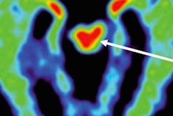
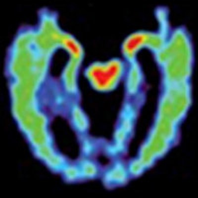
 FDDNP-PET images show a normal brain scan (left), a patient with suspected CTE (middle), and an Alzheimer's patient (right). Red and yellow indicate more abnormal brain proteins (tau and amyloid). The suspected person with CTE has high levels of FDDNP signal in the midbrain (red central area) and amygdalae (smaller red areas). In this more advanced case of suspected CTE, the FDDNP signal (yellow/green) is seen throughout the cortex, which is where most of the FDDNP signal is located in the Alzheimer's patient. Images courtesy of PNAS and David Geffen School of Medicine at UCLA.
The FDDNP-PET scans revealed that scans of the retired football players showed tau deposit patterns consistent with patterns found in autopsies of people with CTE.
FDDNP-PET images also illustrated different brain imaging patterns of concussed individuals compared with scans of healthy subjects and Alzheimer's patients. The former athletes also had higher levels of FDDNP in the amygdala and subcortical regions of the brain -- which control learning, memory, behavior, emotions, and other mental and physical functions -- than healthy people and Alzheimer's patients.
Barrio and colleagues speculated that the results could help better identify brain disorders in athletes and help with the development and testing of treatments to delay the progression of CTE before significant brain damage and symptoms can occur.
UCLA researchers now plan to add more clinical sites to advance their study and will follow subjects over time to determine how effectively FDDNP can detect possible CTE and predict future symptoms.
Barrio and several colleagues also have formed a company called TauMark, to which they have licensed the intellectual property of FDDNP and its use with PET. Barrio, Dr. Gary Small, and Dr. Sung-Cheng Huang are co-inventors of the PET marker. Barrio and Small have a financial interest in TauMark.